Dengue virus antibodies may worsen a Zika infection

By Sharon Isern, Florida Gulf Coast University.

The World Health Organization declared in November that Zika was no longer a public health emergency of international concern.

That doesn’t mean concern over Zika is over, but now that a link between Zika and microcephaly has been established, it is viewed as a long-term problem, which requires constant attention.

While researchers have concluded that Zika infection can cause microcephaly and other birth defects such as eye damage in newborns, there are still many unanswered questions about the virus.

Earlier Zika outbreaks in Africa and Asia were gradual, continuous and associated with mild clinical outcomes, but the Zika outbreaks in the Pacific in 2013 and 2014 and the Americas in 2015 and 2016 have been explosive. They have been associated with severe disease, including birth defects in newborns and Guillain-Barre, a condition that can cause temporary paralysis in adults. Scientists are trying to figure out why.

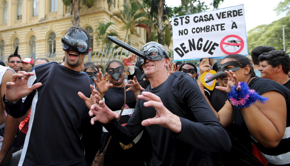
I study flaviviruses, which include Zika and dengue, at Florida Gulf Coast University. Like other flavivirus researchers, I am turning to dengue to better understand Zika. Dengue, a close relative of Zika, is regularly found in places like Brazil, and is spread by the same mosquitoes, Aedes aegypti.

My colleagues and I wanted to find out whether having immunity to dengue from an earlier infection could make a Zika infection worse.

Aedes aegypti mosquitoes can transmit both Zika and dengue viruses.
AP Photo/Felipe Dana, File

A brief history of Zika

Zika was first isolated in Uganda in 1947. For decades Zika infections in humans were sporadic. Perhaps cases of Zika went underreported, since its symptoms were similar to other fever-causing diseases and most cases are asymptomatic.

By the 1980s, Zika had spread beyond Africa and had become endemic, or habitually present, in Asia. Many individuals living in these regions may be immune to the virus.

The first reported Zika outbreak outside of Africa and Asia occurred in the Pacific, in Micronesia, in 2007. To our knowledge there were no associations with microcephaly or Guillain-Barre reported at the time.

Zika transmission in the Pacific wasn’t reported again until 2013, when French Polynesia experienced an explosive outbreak. In 2014 further outbreaks were reported in New Caledonia, Easter Island and the Cook Islands. When French Polynesia experienced another outbreak in 2014, there were reports of Zika being transmitted to babies, most likely in utero, and complications associated with Guillian-Barre in adults.

Map showing countries affected by the Zika virus. Reuters

By early 2015 the virus had spread to the Americas, and the first confirmed case of locally acquired Zika in the region was confirmed in May 2015 by Brazil’s National Reference Laboratory. The Pan American Health Organization reports that Zika virus transmission had occurred in over 48 countries or territories in the Americas, with 177,614 confirmed cases of locally acquired Zika in the region, over half a million suspected cases, and 2,525 confirmed congenital syndrome associated cases with Zika infections as of Dec. 29.

Why did Zika start to cause explosive outbreaks? And why did it start to cause more health problems?

Dengue a common connector

A few factors might explain. For instance, perhaps the difference may lie in the age of the person exposed to Zika.

If children are infected before they reach puberty, they become immune and cannot pass Zika along to their children. And in parts of the world where Zika is endemic, people are more likely to have been exposed and become immune while young.

The scenario we’ve seen in the Americas is different. Since Zika had not been reported in the region prior to 2015, people, including women of child-bearing age, had never been exposed to the virus. However, this doesn’t explain why certain people develop severe disease, whereas others do not.

Dengue virus is endemic many parts of Asia, Africa and the Americas, infecting up to 100 million people globally each year. And the areas of the Pacific and the Americas that experienced explosive Zika outbreaks have two things in common: they had not been exposed to Zika before and dengue is endemic. And dengue may provide a clue to why Zika has caused severe disease in these new outbreaks.

Could a prior dengue infection make Zika worse?

When a person is infected with a particular virus for the first time, the immune system springs into action, producing antibodies to destroy it. The next time a person encounters that virus, the body produces those antibodies again to fight back, preventing illness. This is called immunity.

There are four different kinds of dengue virus, called serotypes. Antibodies produced during infection with one dengue serotype confer lifelong immunity against only that particular serotype. If a person is infected with another serotype later on, the antibodies from the earlier infection will bind to the new virus type, but can’t prevent it from infecting cells.

Instead, the bound antibodies can transport the viruses to immune cells that are normally not infected by dengue. In other words, if a person is infected with one serotype of dengue and then gets infected with another serotype, the antibodies from the previous infection then make the new serotype infect cells that it otherwise wouldn’t. Then the virus can reproduce to very high numbers in these cells, leading to severe disease. This process is called antibody-dependent enhancement, or ADE.

Zika virus is closely related to dengue and has been shown to undergo ADE in response to other flavivirus antibodies. Zika virus antibodies in turn have been shown to have a similar effect on related viruses. And other researchers have shown that preexisting immunity to Zika can enhance dengue virus disease severity in animals.

A digitally colorized electron microscope from the CDC shows the Zika virus, in red. Cynthia Goldsmith/CDC via AP

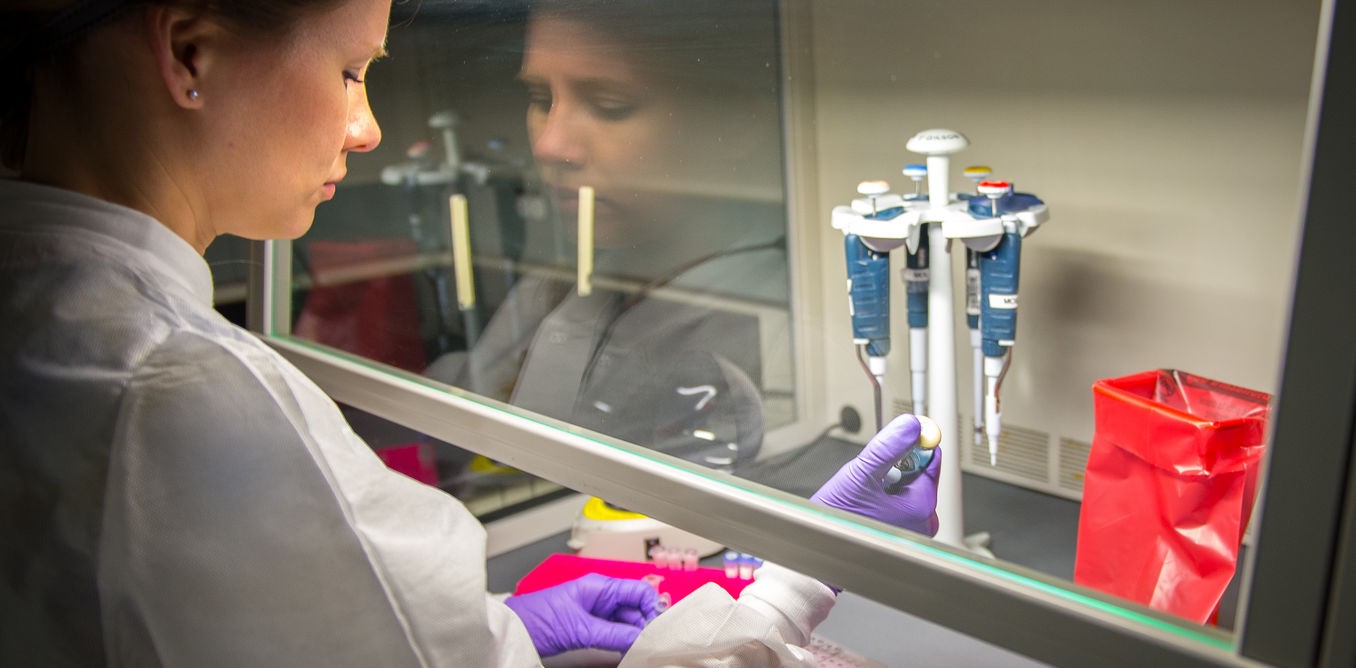
My lab studied the African strain of the Zika virus in 2015 before the connection with microcephaly was known.

Our results, posted in April to bioRxiv, a preprint server for biology, showed that antibodies from a prior dengue virus infection greatly enhanced Zika virus production in cell culture. Other groups have since independently verified our work. And, as we found in a more recent study, the same results hold true with a strain of Zika isolated in Puerto Rico by the CDC.

Our findings suggest that preexisting dengue virus antibodies may enhance Zika virus infection in patients, potentially making the infection more severe.

However, this correlation needs to be confirmed in the clinic. We do not know how many people infected with Zika in the outbreaks in the Pacific and in the Americas had prior dengue infections. But since the virus is endemic in those areas, it is possible that some people may have been infected with dengue before they were infected with Zika.

Zika’s spread may become limited

Like dengue and other mosquito-borne viruses, Zika spread is seasonal, and outbreaks occur when mosquitoes are abundant. As the United States heads into winter and South America heads into summer, what can we expect with regard to Zika and dengue and disease severity?

As more and more people acquire immunity to Zika, its spread will be limited to those who have never been exposed. Given enough time, new infections will be limited to the young. As long as infections occur prior to child-bearing years, microcephaly in newborns should not be a concern.

If these studies hold true in the clinic, cocirculation of Zika and dengue could increase the severity of both viruses. Vaccines to protect against Zika, and dengue for that matter, would need to be designed carefully so that vaccination with one does not enhance a natural infection with the other virus.

Public health emergency or not, there is still much to be learned about Zika virus and its associated consequences.

The ConversationSharon Isern, Professor of Biological Sciences, Florida Gulf Coast University

This article was originally published on The Conversation. Read the original article.

Now, Check Out:

Why you can’t fry eggs (or testicles) with a cellphone

By Timothy J. Jorgensen, Georgetown University.

A minor craze in men’s underwear fashions these days seems to be briefs that shield the genitals from cellphone radiation. The sales claim is that these products protect the testicles from the harmful effects of the radio waves emitted by cellphones, and therefore help maintain a robust sperm count and high fertility. These undergarments may shield the testicles from radiation, but do male cellphone users really risk infertility?

The notion that electromagnetic radiation in the radio frequency range can cause male sterility, either temporary or permanent, has been around for a long time. As I describe in my book “Strange Glow: The Story of Radiation,” during World War II some enlisted men would consistently and inexplicably volunteer for radar duty just prior to their scheduled leave days. It turned out that a rumor had been circulating that exposure to radio waves from the radar equipment produced temporary sterility, which the soldiers saw as an employment benefit.

The military wanted to know whether there was any substance to the sterility rumor. So they asked Hermann Muller – a geneticist who won the Nobel Prize for showing that x-rays could cause sterility and genetic mutations – to evaluate the effects of radio waves in the same fruit fly experimental model he had used to show that x-rays impaired reproduction.

Muller could find no dose of radio waves that produced either sterility or genetic mutations, and concluded that radio waves did not present the same threat to fertility that x-rays did. Radio waves were different. But why? Aren’t both x-rays and radio waves electromagnetic radiation?

The electromagnetic spectrum, tiny wavelengths on the left, longer wavelengths on the right.
Inductiveload, CC BY-SA

Yes, they are – but they differ in one key factor: They have very different wavelengths. All electromagnetic radiation travels through space as invisible waves of energy. And it’s the specific wavelength of the radiation that determines all of its effects, both physical and biological. The shorter wavelengths carry higher amounts of energy than the longer wavelengths.

X-rays are able to damage cells and tissues precisely because their wavelengths are extremely short – one-millionth the width of a human hair – and thus are highly energetic and very harmful to cells. Radio waves, in contrast, carry little energy because their wavelengths are very long – about the length of a football field. Such long-wavelength radiations have really low energies – too low to damage cells. And it’s this big difference between the wavelengths of x-rays and radio waves that the infertility theorists fail to recognize.

X-rays, and other high-energy waves, produce sterility by killing off the testicular cells that make sperm – the “spermatogonia.” And x-ray doses must be extremely high to kill enough cells to produce sterility. Still, even when the doses are high, the sterility effect is usually temporary because the surviving spermatogonia are able to spawn replacements for their dead comrades, and sperm counts typically return to their normal levels within a few months.

So, if high doses of highly energetic x-rays are needed to kill enough cells to produce sterility, how can low doses of radio waves with energies too low to kill cells do it? Good question.

Don’t fall for the phone-cooking-egg hoax.

At this point you may be thinking that you’ve seen videos of cellphones cooking eggs. And you’ve even experienced your cellphone getting pretty warm when it’s used heavily. But this doesn’t show that cellphones put out a lot of radiation energy. The cooked egg video is a prank, and the phone gets hot because of the heat generated by the chemical reactions going on within the battery, not from radio waves.

Still you protest: What about those sporadic reports claiming that cellphones suppress sperm counts? For the moment, that’s all they are – sporadic reports, unconfirmed by other investigators. You can find all kinds of random assertions about the effects of radiation on health, both good and bad, most of which imply that there is some type of validated scientific evidence to support the claim. Why not believe all of them?

If we’ve learned anything over the years about scientific evidence, it’s that isolated findings from individual labs, reporting limited experimental data, do not a strong case make. Most of the very limited “scientific” reports of infertility caused by cellphones, often cited by anti-cellphone activists, come from outside the radiation biology community, and are published in lower-tier journals of questionable quality. Few, if any, of these reports make any attempt at actually measuring the radiation doses received from the cellphones (probably because they lack either the expertise or the equipment required to do it).

Human sperm, unconcerned by what’s in your pocket.
Doc. RNDr. Josef Reischig, CSc., CC BY-SA

And none actually measure fertility rates – the health endpoint of concern – but rather measure sperm counts and other sperm quality parameters and then infer that there will be an impact on fertility. In fact, sperm counts can vary widely between normally fertile individuals and even within the same individual from day to day. For example, men who frequently ejaculate have lower sperm counts, as you might expect, because they are regularly jettisoning sperm. (Men who ejaculate daily can have sperm counts 50 percent lower than men who don’t.) Perhaps the allegedly lower sperm counts of cellphone users just means that they are having more sex!

But seriously, the point is this: There are so many things that can affect sperm counts in big ways that minor fluctuations in sperm counts have no practical impact on whether a man will produce babies, even if it were true that cellphones can modestly suppress sperm counts.

It is clear that these infertility claims are not the consensus of the mainstream scientific community – a community that demands more rigorous evidence. There are many excellent laboratories around the world that study radiation effects, and it isn’t difficult to study infertility in fruit flies, mice and even people. (It’s fairly easy to find men willing to donate sperm samples.) If the sterility story were true, there would be a chorus of well-respected laboratories from around the world singing the cellphone infertility song, not just a few.

Guglielmo Marconi, inventor of the radio. Smithsonian Institution

The fact is, the current data suggesting that cellphones cause infertility are too weak to challenge the dogma of over 100 years of commercial experience with radio waves. Radio waves are not unique to cellphones. They have been used for telecommunication ever since Marconi first demonstrated in 1901 that they could carry messages across the entire Atlantic Ocean. Early radio workers received massive doses of radio waves, yet there is no indication they had any problems with their fertility. If they didn’t experience fertility problems with their high doses, how can the relatively low doses from cellphones have such an effect? Hard to understand.

Nevertheless, people can spend their money as they please and wear any underwear they want. But if you are still concerned about radio waves affecting your fertility, why not just carry your cellphone in your shirt pocket rather than your pants, and let your testicles be?

The ConversationTimothy J. Jorgensen, Director of the Health Physics and Radiation Protection Graduate Program and Associate Professor of Radiation Medicine, Georgetown University

This article was originally published on The Conversation. Read the original article.

Now, Check Out:

With legal pot comes a problem: How do we weed out impaired drivers?

By Igor Grant, University of California, San Diego.

On Nov. 8 voters in California, Maine, Massachusetts and Nevada approved ballot measures to legalize recreational cannabis. It is now legal in a total of eight states. And this creates potential problems for road safety. How do we determine who’s impaired and who’s not?

The effects of alcohol vary based on a person’s size and weight, metabolism rate, related food intake and the type and amount of beverage consumed. Even so, alcohol consumption produces fairly straightforward results: The more you drink, the worse you drive. Factors like body size and drinking experience can shift the correlation slightly, but the relationship is still pretty linear, enough to be able to confidently develop a blood alcohol content scale for legally determining drunk driving. Not so with marijuana.

We have a reliable and easy-to-use test to measure blood alcohol concentration. But right now we don’t have a fast, reliable test to gauge whether someone is too doped up to drive.

The need is urgent. The 2014 National Survey on Drug Use and Health reported that 10 million Americans said they had driven while under the influence of illicit drugs during the previous year. Second to alcohol marijuana is the drug most frequently found in drivers involved in crashes.

But how do you know when you’re too stoned to drive? How can police tell?

My colleagues and I at the Center for Medicinal Cannabis Research at UC San Diego have received a US$1.8 million grant from the state of California to gather data about dosages, time and what it takes to impair driving ability – and then create a viable roadside sobriety test for cannabis.

A man smokes a marijuana joint at a party celebrating weed on April 20, 2016, in Seattle.
AP photos/Elaine Thompson

Testing for marijuana isn’t like a BAC test

Alcohol and marijuana both affect mental function, which means they can both impair driving ability.

Some elements of cannabis use are similar. Potency of strain affects potency of effect. Marijuana and its active ingredient – THC – alter brain function, affecting processes like attention, perception and coordination, which are necessary for a complex behavior like driving a car.

Regular users tend to become accustomed to the drug, particularly in terms of cognitive disruption or psycho-motor skills. Because they are accustomed to the drugs’ effects, this means they may function better relative to naïve users.

Smoked marijuana produces a rapid spike in THC concentrations in the blood, followed by a decline as the drug redistributes to tissues, including the brain. The psychological impact depends upon a host of variables.

Let’s say, for example, a person smokes a joint and gets into his car. THC levels in his blood are likely to be quite high, but his cognitive functions and driving skills may not yet be impaired because the drug hasn’t yet significantly impacted the brain. But another driver might use cannabis but wait a few hours before getting behind the wheel. Her THC blood levels are now quite low, but she’s impaired because drug concentrations remain high in her brain.

Six states have set limits for THC in drivers’ blood, and nine other states have zero-tolerance laws, making the presence of THC in the drivers blood illegal.

But unlike alcohol, evidence of cannabis use can linger long after its effects have worn off, particularly if people are regular users or consume a lot in a single episode. Among chronic users, it may not clear out of their systems for weeks. Therefore, unlike blood alcohol concentration, the presence and amount of different cannabis compounds in the blood or urine do not necessarily tell you whether the driver is impaired due to marijuana.

This is why a quick and simple assessment of whether someone is driving while under the influence is difficult. And that is a necessity for any type of effective roadside sobriety test.

To create a fast and easy-to-use test, there are a few questions about marijuana that our team at UC San Diego has to answer.

How high is too high to drive?
Ignition key image via www.shutterstock.com.

How much marijuana is too much to drive?

Current blood, breath, saliva and urine tests have been challenged as unreliable in court, though they are used to prove that someone has ingested marijuana.

In California and elsewhere, the primary assessment of impairment is the law enforcement officer’s field sobriety test.

One specific challenge is determining the relationship of dose or potency, and time since consumption, to impairment. While there has been some research in this area, the studies have not comprehensively examined the issues of dose and time course of impairment. The lack of data is one of the big reasons for our work now.

Later this year, we will begin controlled experiments in which participants will smoke varying amounts of cannabis in varying strengths and then operate a driving simulator. We’ll look for impairment effects in the immediate period after exposure and over subsequent hours.

We’ll also investigate the relationship between THC and other cannabinoid levels in blood to different measures, such as saliva or exhaled breath. Roadside blood sampling is impractical, but perhaps there is an easier, reliable indicator of marijuana exposure.

Finally, there is the goal of finding the best way to assess impairment. A driver suspected of being high might be asked to follow with his finger a square moving around on a device’s screen, a test of critical tracking. Or she might perform tablet tests that more validly simulate the demands of driving.

The idea is to determine whether and how these measures – drug intake, biomarkers, objective cognitive performance and driving ability – correlate to produce an evidence-based, broadly applicable assessment standard and tool.

The ConversationIgor Grant, Professor and Chair of Department of Psychiatry and Director, Center for Medical Cannabis Research, University of California, San Diego

This article was originally published on The Conversation. Read the original article.

Now, Check Out:

This Newly-Identified Human Antibody May Lead to Zika Vaccination

Researchers have identified a human antibody that prevents Zika from infecting the fetus and damaging the placenta in pregnant mice. The antibody also protects adult mice from Zika disease.

The most devastating consequence of Zika virus infection is the development of microcephaly, or an abnormally small head, in fetuses infected in utero.

“This is the first antiviral that has been shown to work in pregnancy to protect developing fetuses from Zika virus,” says Michael Diamond, professor of medicine at Washington University School of Medicine in St. Louis and the study’s co-senior author. “This is proof of principle that Zika virus during pregnancy is treatable, and we already have a human antibody that treats it, at least in mice.”

ZIKV-117

Diamond, co-senior author James Crowe Jr. of Vanderbilt, and colleagues screened 29 anti-Zika antibodies from people who had recovered from Zika infection. They found one, called ZIKV-117, that efficiently neutralized in the lab five Zika strains—representing the worldwide diversity of the virus.

To test whether the antibody also protects living animals, the researchers gave the antibody to pregnant mice either one day before or one day after they were infected with the virus. In both cases, antibody treatment markedly reduced the levels of virus in pregnant females and their fetuses, as well as in the placentas, compared with pregnant mice that did not get the antibody.

“These naturally occurring antibodies isolated from humans represent the first medical intervention that prevents Zika infection and damage to fetuses,” Crowe says.

The placentas from the treated females appeared normal and healthy, unlike those from the untreated females, which showed destruction of the placental structure. Damage to the placenta can cause slow fetal growth and even can cause fetal death, both of which are associated with Zika infection in humans.

“We did not see any damage to the fetal blood vessels, thinning of the placenta, or any growth restriction in the fetuses of the antibody-treated mice,” says coauthor Indira Mysorekar, an associate professor of obstetrics and gynecology, and of pathology and immunology at Washington University, and co-director of the university’s Center for Reproductive Sciences.

“The anti-Zika antibodies are able to keep the fetus safe from harm by blocking the virus from crossing the placenta.”

The antibody also protected adult male mice against a lethal dose of Zika virus, even when given five days after initial infection. Zika is rarely lethal in humans, so using a lethal dose allowed the scientists to see how well the antibody works under the most stringent conditions.

“We stacked the deck against ourselves by using a highly pathogenic strain of Zika, and even in that case, the antibody protected the mice,” says Diamond, who is also a professor of pathology and immunology, and of molecular microbiology.

Support for a vaccine

These findings provide evidence that antibodies alone can protect adults and fetuses from Zika. Further, they suggest that a vaccine that elicits protective antibodies in women also may protect their fetuses in current and future pregnancies. A vaccine is already in human trials, but it was never tested in pregnant animals, so this new study represents strong evidence that a vaccine that elicits protective antibodies in adults is likely to protect fetuses as well.

A Zika vaccine is likely to be the cheapest and simplest method of preventing Zika-related birth defects. However, there is an outside possibility that a Zika vaccine could worsen symptoms in people who encounter the virus later. This is known to occur with dengue virus, a close relative of Zika. People who have antibodies against one strain of dengue virus get sicker when infected with a second strain than those who do not have such antibodies.

The phenomenon, known as antibody-dependent enhancement, has been observed with Zika in a petri dish but never in living animals or in epidemiologic surveys of people in Zika-endemic regions.

Nonetheless, the researchers tested whether they could eliminate the possibility of antibody-dependent enhancement of Zika infection by modifying the antibody so it could not participate in the process. The modified antibody, they showed, was just as effective as the original at protecting the placenta and fetus.

Treatment during pregnancy?

Until a human vaccine is available, it may be possible to protect fetuses by administering antibodies to pregnant women in an attempt to prevent transmission from mother to fetus. Under this scenario, a woman living in a Zika-endemic area would receive the antibodies throughout her pregnancy, starting when she first learns she is pregnant, regardless of whether she is diagnosed with Zika. Alternatively, pregnant women or their partners with acute infection could be treated with antibodies.

Crowe is continuing the process of developing the antibody as a potential therapeutic, ramping up production and laying the groundwork for human studies. Meanwhile, Diamond is focusing on determining whether antibodies could be used to clear persistent Zika infection. Together, they are working with others to gain a higher-resolution understanding of how ZIKV-117 binds the virus and inhibits infection.

“We know that Zika can persist in certain parts of the body, such as the eyes and the testes, where it can cause long-term damage, at least in mice,” Diamond says. “We showed that the antibody can prevent disease, and now we want to know whether it can clear persistent infection from those parts of the body.”

The study appears online in Nature.

Source: Republished from Futurity.org as a derivative work under the Attribution 4.0 International license. Original article posted to Futurity by .

Now, Check Out:

The next frontier in medical sensing: Threads coated in nanomaterials

By Sameer Sonkusale, Tufts University.

Doctors have various ways to assess your health. For example, they measure your heart rate and blood pressure to indirectly assess your heart function, or straightforwardly test a blood sample for iron content to diagnose anemia. But there are plenty of situations in which that sort of monitoring just isn’t possible.

To test the health of muscle and bone in contact with a hip replacement, for example, requires a complicated – and expensive – procedure. And if problems are found, it’s often too late to truly fix them. The same is true when dealing with deep wounds or internal incisions from surgery.

In my engineering lab at Tufts University, we asked ourselves whether we could make sensors that could be seamlessly embedded in body tissue or organs – and yet could communicate to monitors outside the body in real time. The first concern, of course, would be to make sure that the materials wouldn’t cause infection or an immune response from the body. The sensors would also need to match the mechanical properties of the body part they would be embedded in: soft for organs and stretchable for muscle. And, ideally, they would be relatively inexpensive to make in large quantities.

Our search for materials we might use led us to a surprising candidate – threads, just like what our clothes are made of. Thread has many advantages. It is abundant, easy to make and very inexpensive. Threads can be made flexible and stretchable – and even from materials that aren’t rejected by the body. In addition, doctors are very comfortable working with threads: They routinely use sutures to stitch up open wounds. What if we could embed sensor functions into threads?

Finding the right sensor

Today’s medical sensors are typically rigid and flat – which limits them to monitoring surfaces such as the scalp or skin. But most organs and tissues are three-dimensional heterogeneous multilayered biological structures. To monitor them, we need something much more like a thread.

Nanomaterials can be organic or inorganic, inert or bioactive, and can be designed with physical and chemical properties that are useful for medical sensing. For example, carbon nanotubes are amazingly versatile – their electrical conductivity can be customized, which has led to them being the basis of the next generation of sensors and electronic transistors. They can even detect single molecules of DNA and proteins. The organic nanomaterial polyaniline has a similarly broad range of applications, notably its conductivity depends on the strength of the acid or base it is in contact with.

Making the materials

To make sensing threads, we start with cotton and other conventional threads, dip them in liquids containing different nanomaterials, and rapidly dry them. Depending on the properties of the nanomaterial we use, these can monitor mechanical or chemical activity.

For example, coating stretchable rubber fiber with carbon nanotubes and silicone can make threads that can sense and measure physical strain. As they stretch, the threads’ electrical properties change in ways we can monitor externally. This can be used to monitor wound healing or muscle strain experienced due to artificial implants. After an implant, abnormal strain could be a sign of slow healing, or even improper placement of the device. Threads monitoring strain levels can send a message to both patient and doctor so that treatment can be modified appropriately.

Monitoring the electricity flow between one cotton thread coated with carbon nanotubes and polyaniline nanofibers, and another coated with silver and silver chloride, allows us to measure acidity, which can be a sign of infection.

To help people who need to monitor their blood sugar levels, we can coat a thread with glucose oxidase, which reacts with glucose to generate an electrical signal indicating how much sugar is in the patient’s blood. Similarly, coating conductive threads with other nanomaterials sensitive to specific elements or chemicals can help doctors measure potassium and sodium levels or other metabolic markers in your blood.

Multiple uses

Beyond sensing abilities, many thread materials, such as cotton, have another useful property: wicking. They can move liquid along their length via capillary action without needing a pump, the way melted wax flows up a candlewick to feed the flame.

Liquid flowing in threads sutured into skin.
Nano Lab, Tufts University, CC BY-ND

We used cotton threads to transport interstitial fluid, which fills in the gaps between cells, from the places it normally exists toward sensing threads located elsewhere. The sensing threads send their electronic signals to an external device housed in a flexible patch, along with a button battery and a small antenna. There, the signals are amplified, digitized and transmitted wirelessly to a smartphone or any Wi-Fi connected device.

These transport-sensing measuring-transmission systems are so small that they can be powered with a tiny battery sitting on top of the skin or could get energy from glucose in the patient’s blood. That could allow doctors to keep a continuous eye on patients’ health remotely and unobtrusively.

Smart threads can monitor wounds using a suite of physical and chemical sensors made using threads and passing information to a skin-surface transmitter.
Nano Lab, Tufts University, CC BY-ND

This type of integrated, wireless monitoring has several advantages over current systems. First, the patient can move around freely, rather than being confined to a hospital bed. In addition, real-time data-gathering provides much more accurate information than periodic testing at a hospital or doctor’s office. And it reduces the cost of health care by moving treatment, monitoring and diagnosis out of the hospital.

So far our testing of nano-infused threads has been in sterile lab environments in rodents. The next step is to perform more tests in animals, particularly to monitor how well the threads do in living tissue over long periods of time. Then we’d move toward testing in humans.

Now that we’ve begun exploring the possibilities of threads, potential uses seem to be everywhere. Diabetic patients can have trouble with wounds resisting healing, which can lead to infection, and even amputation. A few choice stitches using sensing threads could let doctors detect these problems at extremely early stages – much sooner than we can today – and take action to prevent them from worsening. Sensing threads can even be woven into bandages, wound dressings or hospital bed sheets to monitor patients’ progress, and raise alarms before problems get out of control.

The ConversationSameer Sonkusale, Professor of Electrical and Computer Engineering, Tufts University

This article was originally published on The Conversation. Read the original article.

Now, Check Out:

Yoshinori Ohsumi – a deserving winner of the Nobel Prize for physiology or medicine

By David Rubinsztein, University of Cambridge.

I am delighted that Yoshinori Ohsumi won this year’s Nobel Prize in physiology or medicine. His pioneering work in yeast led to the discovery of genes and biological processes that are needed for autophagy.

Autophagy (from the Greek for “self-eating”) is the mechanism by which cells break down and recycle cellular content. Without this vital housekeeping role we’d be more prone to cancer, Parkinson’s and other age-related disorders.

Although scientists have been aware of autophagy since the 1960s, it wasn’t until Ohsumi’s experiments with yeast in the 1990s that we began to understand the important role of this biological process.

The autophagy process is remarkably similar across lifeforms. One function that is the same, from yeast to humans, is to protect cells against starvation and related stresses. In these conditions, autophagy allows cells to degrade large molecules into basic building blocks, which are used as energy sources. The discovery of key yeast autophagy genes that was led by Ohsumi was particularly powerful because it helped scientists to quickly identify the genes in mammals that have similar functions. This, in turn, has provided vital tools for laboratories around the world to study the roles of autophagy in human health and disease.

With the knowledge that various mammalian genes are needed for autophagy, researchers could then remove these genes from cells or animals, including mice, and examine their functions. These types of studies have highlighted the importance of autophagy in processes including infection and immunity, neurodegenerative diseases and cancer.

The importance of Ohsumi’s findings

My laboratory, for example, found that autophagy can break down the proteins responsible for various neurological diseases, including forms of dementia (caused by tau), Parkinson’s disease (alpha-synuclein) and Huntington’s disease (mutant huntingtin). We are pursuing the idea that by increasing the autophagy process we could potentially treat some of these conditions.

A tau protein fragment. molekuul_be/Shutterstock.com

Another important consequence of Ohsumi’s discoveries is that they allowed subsequent studies that aimed to understand the mechanisms by which autophagy proteins actually control this process. Indeed, Ohsumi’s group have also made seminal contributions in this domain.

This Nobel prize highlights some other key characteristics of Ohsumi and his work. One is that his laboratory works on yeast. At the time he made his discoveries in the 1990s, no one would have guessed that they would have such far-reaching implications for human health. Essentially, he was studying autophagy in yeast because he was curious. This basic research yielded the foundation for an entire field, which has grown rapidly in recent years, especially as its relevance for health has become more apparent. This should serve as a reminder to those influencing science strategy that groundbreaking discoveries are often unexpected and that one should not only support science where the endpoint appears to be obviously relevant to health.

Ohsumi has also nurtured outstanding scientists like Noboru Mizushima and Tamotsu Yoshimori, who have been major contributors to the understanding of autophagy in mammals. Perhaps most importantly, he continues to do interesting and fundamental work. This Nobel prize is very well deserved for the man who opened the door to an important field.

The ConversationDavid Rubinsztein, Professor of molecular neurogenetics, University of Cambridge

This article was originally published on The Conversation. Read the original article.

Now, Check Out:

Fungus is Found to be Key Factor in Crohn’s Disease

A Case Western Reserve University School of Medicine-led team of international researchers has for the first time identified a fungus as a key factor in the development of Crohn’s disease. The researchers also linked a new bacterium to the previous bacteria associated with Crohn’s. The groundbreaking findings, published on September 20th in mBio, could lead to potential new treatments and ultimately, cures for the debilitating inflammatory bowel disease, which causes severe abdominal pain, diarrhea, weight loss, and fatigue.

“We already know that bacteria, in addition to genetic and dietary factors, play a major role in causing Crohn’s disease,” said the study’s senior and corresponding author, Mahmoud A Ghannoum, PhD, professor and director of the Center for Medical Mycology at Case Western Reserve and University Hospitals Cleveland Medical Center “Essentially, patients with Crohn’s have abnormal immune responses to these bacteria, which inhabit the intestines of all people. While most researchers focus their investigations on these bacteria, few have examined the role of fungi, which are also present in everyone’s intestines. Our study adds significant new information to understanding why some people develop Crohn’s disease. Equally important, it can result in a new generation of treatments, including medications and probiotics, which hold the potential for making qualitative and quantitative differences in the lives of people suffering from Crohn’s.”

Both bacteria and fungi are microorganisms — infinitesimal forms of life that can only be seen with a microscope. Fungi are eukaryotes: organism whose cells contain a nucleus; they are closer to humans than bacteria, which are prokaryotes: single-celled forms of life with no nucleus. Collectively, the fungal community that inhabits the human body is known as the mycobiome, while the bacteria are called the bacteriome. (Fungi and bacteria are present throughout the body; previously Ghannoum had found that people harbor between nine and 23 fungal species in their mouths.)

The researchers assessed the mycobiome and bacteriome of patients with Crohn’s disease and their Crohn’s-free first degree relatives in nine families in northern France and Belgium, and in Crohn’s-free individuals from four families living in the same geographic area. Specifically, they analyzed fecal samples of 20 Crohn’s and 28 Crohn’s-free patients from nine families and of 21 Crohn’s-free patients of four families. The researchers found strong fungal-bacterial interactions in those with Crohn’s disease: two bacteria (Escherichia coli and Serratia marcescens) and one fungus (Candida tropicalis) moved in lock step. The presence of all three in the sick family members was significantly higher compared to their healthy relatives, suggesting that the bacteria and fungus interact in the intestines. Additionally, test-tube research by the Ghannoum-led team found that the three work together (with the E. coli cells fusing to the fungal cells and S. marcescens forming a bridge connecting the microbes) to produce a biofilm — a thin, slimy layer of microorganisms found in the body that adheres to, among other sites, a portion of the intestines — which can prompt inflammation that results in the symptoms of Crohn’s disease.

This is first time any fungus has been linked to Crohn’s in humans; previously it was only found in mice with the disease. The study is also the first to include S. marcescens in the Crohn’s-linked bacteriome. Additionally, the researchers found that the presence of beneficial bacteria was significantly lower in the Crohn’s patients, corroborating previous research findings.

“Among hundreds of bacterial and fungal species inhabiting the intestines, it is telling that the three we identified were so highly correlated in Crohn’s patients,” said Ghannoum. “Furthermore, we found strong similarities in what may be called the ‘gut profiles’ of the Crohn’s-affected families, which were strikingly different from the Crohn’s-free families. We have to be careful, though, and not solely attribute Crohn’s disease to the bacterial and fungal makeups of our intestines. For example, we know that family members also share diet and environment to significant degrees. Further research is needed to be even more specific in identifying precipitators and contributors of Crohn’s.”


Source: News release on Science Daily

Featured Photo Credit: © vaakim / Fotolia

Now, Check Out:

Feed a virus but starve bacteria? When you’re sick, it may really matter

By Ruslan Medzhitov, Yale University.

Think back to the last time you came down with a cold and what it felt like to be sick. For most people, the feeling of sickness is a set of psychological and behavioral changes including fatigue, lethargy, changes in appetite, changes in sleep patterns and a desire to be away from others.

Of course, none of these changes feel particularly good, but what if they are actually good for us in terms of recovering from the infection?

Interestingly, these infection-induced behavioral changes, collectively known as “sickness behaviors,” occur in most other animals – from your pet dogs and cats to the worms in your backyard. Because so many animals exhibit sickness behaviors during infection, scientists have thought for decades that these behaviors may protect us from infections.

In our immunobiology lab at Yale University, we are interested in these sickness behaviors and most recently have focused on the aspect of appetite loss during infection. If all sickness behaviors indeed help us survive infections, then how does loss of appetite specifically fit in?

One common theory is that although we are starving ourselves, starvation is worse for the bacteria or virus than it is for us. Some scientific evidence supports this theory, but a lot does not.

Recently we ventured to reexamine why we lose our appetites when we get sick.

Why your appetite matters when you get an infection

The question of whether or not we should eat when we get sick is commonly argued, both at home and in the hospital. Every family has its own beliefs about how to address appetite loss during infection.

Some believe it’s best to keep well-fed regardless of desire to eat, some swear by old adages like “feed a fever, starve a cold” and few suggest letting the sick individual’s appetite guide their food consumption. Determining which of these is the right approach – or if it even matters – could help people recover better from mild infections.

Another, perhaps more important, reason to understand appetite changes during infection is to improve survival of critically ill patients in intensive care units across the world. Critically ill patients often cannot feed themselves, so doctors generally feed them during the time of critical illness.

But how much food is the right amount of food? And what type of food is best? And which patients should we feed? Doctors have struggled with these questions for decades and have performed many clinical trials to test different feeding regimens, but no definitive conclusions have been reached.

If we could understand the role of appetite in infection, we could provide more rational care for infected patients at home and in the hospital.

Is losing your appetite a good thing when you’re sick?

Based on our recent findings, it depends.

Like humans, lab mice lose their appetite when infected. When we infected mice with the bacteria Listeria monocytogenes and fed them, they died at a much higher frequency.

In stark contrast, when we infected mice with the flu virus and fed them, they survived better than their unfed counterparts.

Interestingly, these same effects were observed when we substituted live bacteria with only a small component of the bacterial wall or replaced a live virus with a synthetic mimic of a virus component. These components are found in many bacteria and viruses, respectively, suggesting that the opposing effects of feeding that we observed might extend to many bacteria and viruses.

We found the glucose in food was largely responsible for the effects of feeding. These effects were reversed when we blocked the cell’s ability to use glucose with chemicals called 2-deoxy-glucose (2DG) or D-manno-heptulose (DMH).

Why does eating affect bacterial and viral infections differently?

T cells, which fight infection, can also harm other cells. T cells, which fight infection, can also harm other cells.
From www.shutterstock.com

Surviving an infection is a complex process with many factors to consider. During an infection, there are two things that can cause damage to the body. The first is direct damage to the body caused by the microbe. The second is collateral damage caused by the immune response.

The immune system’s early defenses are relatively nonspecific – they can be thought of as grenades rather than sniper rifles. Because of this, the immune system can damage other parts of the body in an effort to clear the infection. To defend against this, tissues in the body have mechanisms to detoxify or resist the toxic agents the immune system uses to attack invaders. The ability of tissues to do this is called tissue tolerance.

In our recent study, we found that tissue tolerance to bacterial and viral infections required different metabolic fuels.

Ketone bodies, which are a fuel made by the liver during extended periods of fasting, help to defend against collateral damage from antibacterial immune responses.

In contrast, glucose, which is abundant when eating, helps to defend against the collateral damage of an antiviral immune response.

What does this mean for humans?

It’s too early to say.

The bottom line is that mice are not people. Many promising treatments in mouse models have failed to translate into people. The concepts we’ve discussed here will need to be confirmed and reconfirmed many times over in humans before they can be applied.

But this study does suggest how we should think about our choice of food during illness. Until now, nutrition selection, especially in the setting of critical illness, was arbitrarily chosen, and mostly selected based on the type of organ failure that the patient had.

Our studies would suggest that what may matter more in selecting nutrition for critically ill patients is what kind of infection they have. As for less serious infections, our work suggests that what you feel like eating when you don’t feel well may be your body’s way of telling you how best to optimize your response to the infection.

So maybe this is what Grandma meant when she told you to “starve a fever, stuff a cold.” Maybe she already knew that different infections required different kinds of nutrition for you to get better quicker. Maybe she knew that if you behaved a certain way, honey tea was best for you, or chicken soup. Maybe Grandma was right? We hope to find out as we work to translate this research to humans.

The ConversationRuslan Medzhitov, Professor of Immunobiology, Yale University

This article was originally published on The Conversation. Read the original article.

Zika virus: Only a few small outbreaks likely to occur in the continental US

By Natalie Exner Dean, University of Florida; Alessandro Vespignani, Northeastern University; Elizabeth Halloran, University of Washington, and Ira Longini, University of Florida.

It is estimated that about 80 percent of Zika infections are asymptomatic or have symptoms so mild that the disease is not detected. This means the number of cases reported by disease surveillance systems in the U.S. and across the world might be only a small fraction of the actual number of infections. In fact, it’s likely we are are underestimating imported cases in the U.S. and even likely some locally spread cases.

In this situation, mathematical and computational models that account for mosquito populations, human mobility, infrastructure and other factors that influence the spread of Zika are valuable because they can generate estimates of the full extent of the epidemic.

This is what our research group, made up of physicists, biostatisticians and computer scientists, has done for Zika. The Global Epidemic and Mobility Model (GLEAM) can model the spread of Zika through countries and geographical regions.

Our model suggests that while more cases of Zika can be expected in the continental U.S., outbreaks will probably be small and are not projected to spread. By contrast, some countries, like Brazil, have already seen widespread outbreaks.

How does the model work?

Zika is primarily transmitted by Aedes mosquitoes. For a mosquito to transmit Zika to a human, it must first have bitten a human infected with the virus. If enough people infected with Zika travel to a new area with these mosquitoes, the virus could spread in a new geographic region.

That means models for Zika transmission need to take factors like mosquito population, human mobility and temperature, among others, into account.

So we begin by dividing the population of the Americas into geographical cells of similar size, and grouping these cells into subpopulations centered around major transportation hubs.
7
Our model also incorporates data on the density of the mosquitoes that transmit Zika, Aedes aegypti and Aedes albopictus, within those subpopulations. Mosquitoes need warm weather to thrive, so we include a daily estimated temperature for each subpopulation. That allows us to factor seasonal temperature changes into our simulations.

To breed, mosquitoes need standing water, and to spread Zika, they need people to feed on. Areas with standing water, fewer window screens and less air conditioning, which are often lower-income areas, are at greater risk. The model uses detailed data about socioeconomics for each subpopulation, as well as data on the relationship between socioeconomic status and risk of exposure to mosquito-borne disease.

Once all of these factors are incorporated into the model, we simulate a Zika outbreak. These simulations are meant to project what will happen next with Zika, so they need to include information about what has already happened. The simulations were calibrated to match data from countries that experienced the epidemic first, like Brazil and Colombia.

We started by “introducing” Zika into one of 12 major transportation hubs in Brazil. Each calibration starts with a different time and place where Zika was first introduced into the country, and simulates about 500,000 possible epidemics. From those we select a few thousand that match surveillance data to project the epidemic forward. Randomness is also incorporated into the simulations so that the resulting “epidemics” can reflect the natural variability in how diseases spread.

Zika’s spread in the U.S. will be limited

Based on current data, our model projects only small outbreaks from mosquito transmission in the continental U.S. that are likely to die out before spreading to new areas.

Alabama, Arkansas, Georgia, Louisiana, Mississippi, Oklahoma, South Carolina and Texas are at risk of these small outbreaks. This is because it is warm enough in these states through the summer and fall to sustain mosquito transmission.

A map of North America plotting the local Zika transmission potential over time and space. Areas displayed in red have the highest Zika transmission potential.
Zhang et al, CC BY-NC-ND

But the median number of daily cases from local mosquito transmission in these states is projected to be zero. This means that in general we do not expect an outbreak to happen, though small outbreaks are possible. Any outbreaks in these states are expected to end by November or December 2016, consistent with declining temperatures and the end of mosquito season.

Florida, on the other hand, may observe sustained transmission between September and November 2016. After calibrating the model with available surveillance data through mid-August, on average, less than 100 symptomatic Zika cases are projected by the second half of September. As many as eight pregnant women could be locally infected in the first trimester, though these women would not give birth until October 2017. In comparison, over 671 pregnant women infected during travel have already been identified in the U.S. as of September 1, 2016. And, as in other states, when mosquito season ends in December, so will Zika transmission from local mosquitoes.

Keep in mind, we are just talking about people getting infected with Zika from local mosquitoes. In the U.S. the number of local cases is expected to be small relative to the number of travel-related infections and to affect comparatively few pregnant women.

The number of travel-related and local cases that are detected by the Zika surveillance system in the continental U.S. is likely much smaller than the total number of infections. Our model estimates that only 2 percent to 5 percent of travel-related infections are detected by surveillance. And local infections may not be detected for individuals without symptoms. But even taking frequent travel-related infections and low detection rates into account, our models project few local cases in the continental U.S.

It’s a different picture for other parts of the Americas. Our models suggest that larger outbreaks occurred or will occur in Brazil, Colombia, Venezuela and Puerto Rico. All have tropical or subtropical climates, have higher densities of the mosquito vectors, and may be at greater risk due to socioeconomic factors.

This is a projection, not a prediction

Remember, these are projections for what might happen, not predictions of what will happen. No model can perfectly replicate reality.

For instance, this model doesn’t account for sexual transmission. We still don’t know how common it is for a person infected with Zika to transmit it during sex. Sexual transmission may proportionally have a larger effect in domestic outbreaks than we realize.

This type of detailed modeling is complex, and that makes it difficult to examine what is happening within states, or even within single counties. It will take more time and data to analyze simulations at such local levels.

Finally, the model does not include any interventions, such as increased mosquito control. Unless other modes of transmission, such as sexual transmission, turn out to be significant factors, our projection might be considered a worst-case scenario.

Model projections like this should be always scrutinized using information about what is happening on the ground. And they need to be recalibrated and refined as new information becomes available.


Computational simulations and projections for the Zika model are a collaboration overseen by the Center for Inference and Dynamics of Infectious Diseases, a Models of Infectious Disease Agent Study Center of Excellence funded by the National Institutes of Health. The collaboration includes Northeastern University, the University of Florida, the Bruno Kessler Foundation, Bocconi University, the Institute for Scientific Interchange Foundation, the Fred Hutchinson Cancer Research Center, and the University of Washington.

The ConversationNatalie Exner Dean, Postdoctoral Associate in Biostatistics, University of Florida; Alessandro Vespignani, Sternberg Family Distinguished University Professor, Northeastern University; Elizabeth Halloran, Researcher, Fred Hutchinson Cancer Research Center and Professor of Biostatistics, University of Washington, and Ira Longini, Professor & Co-director, Center for Statistics and Quantitative Infectious Diseases, University of Florida

This article was originally published on The Conversation. Read the original article.

Now, Check Out:

Why you should dispense with antibacterial soaps

By Sarah Ades, Pennsylvania State University and Kenneth Keiler, Pennsylvania State University.

An FDA ruling on Sept. 2 bans the use of triclosan, triclocarban and 17 other antiseptics from household soaps because they have not been shown to be safe or even have any benefit.

About 40 percent of soaps use at least one of these chemicals, and the chemicals are also found in toothpaste, baby pacifiers, laundry detergents and clothing. It is in some lip glosses, deodorants and pet shampoos.

The current FDA action bans antiseptics like triclosan in household soaps only. It does not apply to other products like antiseptic gels designed to be used without water, antibacterial toothpaste or the many fabrics and household utensils in which antibacterials are embedded. Data suggest that the toothpastes are very effective for people suffering from gum disease, although it is not clear if they provide substantial benefits for those who don’t have gingivitis.

The FDA is currently evaluating the use of antibacterials in gels and will rule on how those products should be handled once the data are in.

Although antibacterials are still in products all around us, the current ban is a significant step forward in limiting their use.

As microbiologists who study a range of chemicals and microbes, we will explain why we don’t we need to kill all the bacteria. We also will explain how antibiotic soaps may even be bad by contributing to antibiotic-resistant strains of bacteria that can be dangerous.

Bacteria can be good

Bacteria are everywhere in the environment and almost everywhere in our bodies, and that is mostly good.

We rely on bacteria in our guts to provide nutrients and to signal our brains, and some bacteria on our skin help protect us from harmful pathogens.

Bacteria in soil can be bad for you. www.shutterstock.com

Some bacteria present in soil and animal waste can cause infections if they are ingested, however, and washing is important to prevent bacteria from spreading to places where they can cause harm.

Washing properly with soap and water removes these potential pathogens. If you have any questions about hand washing, the Centers for Disease Control and Prevention has a great site where you can learn more.

If soap and water are sufficient to remove potential pathogens, why were antibacterials like triclosan and triclocarban added in the first place?

Triclosan was introduced in 1972. These chemicals were originally used for cleaning solutions, such as before and during surgeries, where removing bacteria is critical and exposure for most people is short. Triclosan and triclocarban may be beneficial in these settings, and the FDA ruling does not affect health care or first aid uses of the chemicals.

In the 1990s, manufacturers started to incorporate triclosan and triclocarban in products for the average consumer, and many people were attracted by claims that these products killed more bacteria.

Now antibacterial chemicals can be found in many household products, from baby toys to fabrics to soaps. Laboratory tests show the addition of these chemicals can reduce the number of bacteria in some situations. However, studies in a range of environments, including urban areas in the United States and squatter settlements in Pakistan, have shown that the inclusion of antibacterials in soap does not reduce the spread of infectious disease. Because the goal of washing is human health, these data indicate that antibacterials in consumer soaps do not provide any benefit.

While not all bad, bacteria are promiscuous

What’s the downside to having antibacterials in soap? It is potentially huge, both for those using it and for society as a whole. One concern is whether the antibacterials can directly harm humans.

Triclosan had become so prevalent in household products that in 2003 a nationwide survey of healthy individuals found it in the urine of 75 percent of the 2,517 people tested. Triclosan has also been found in human plasma and breast milk.

Most studies have not shown any direct toxicity from triclosan, but some animal studies indicate that triclosan can disrupt hormone systems. We do not know yet whether triclosan affects hormones in humans.

Another serious concern is the effect of triclosan on antibiotic resistance in bacteria. Bacteria evolve resistance to nearly every threat they face, and triclosan is no exception.

Triclosan isn’t used to treat disease, so why does it matter if some bacteria become resistant? Some of the common mechanisms that bacteria use to evade triclosan also let them evade antibiotics that are needed to treat disease. When triclosan is present in the environment, bacteria that have these resistance mechanisms grow better than bacteria that are still susceptible, so the number of resistant bacteria increases.

Not only are bacteria adaptable, they are also promiscuous. Genes that let them survive antibiotic treatment are often found on pieces of DNA that can be passed from one bacterium to another, spreading resistance.

These mobile pieces of DNA frequently have several different resistance genes, making the bacteria that contain them resistant to many different drugs. Bacteria that are resistant to triclosan are more likely to also be resistant to unrelated antibiotics, suggesting that the prevalence of triclosan can spread multi-drug resistance. As resistance spreads, we will not be able to kill as many pathogens with existing drugs.

Important in some settings

Antibacterial washes are important for surgery. From www.shutterstock.com

Antibiotics were introduced in the 1940s and revolutionized the way we lead our lives. Common infections and minor scrapes that could be fatal became easily treatable. Surgeries that were once unthinkable due to the risk of infection are now routine.

However, bacteria are becoming stronger due to decades of antibiotic use and misuse. New drugs will help, but if we do not protect the antibiotics we have now more people will die from infections that used to be easily treated. Removing triclosan from consumer products will help protect antibiotics and limit the threat of toxicity from extended exposure, without any adverse effect on human health.

The FDA ruling is a welcome first step to cleansing the environment of chemicals that provide little health value to most people but pose significant risk to individuals and to public health. To a large extent, this ruling is a victory of science over advertising.

The ConversationSarah Ades, Associate Professor of Biochemistry and Molecular Biology, Pennsylvania State University and Kenneth Keiler, Professor of Biochemistry and Molecular Biology, Pennsylvania State University

This article was originally published on The Conversation. Read the original article.

Now, Check Out: