Know your bugs – a closer look at viruses, bacteria, and parasites

Arinjay Banerjee, University of Saskatchewan; Jason Byron Perez, University of Saskatchewan, and Simona John von Freyend, Monash University
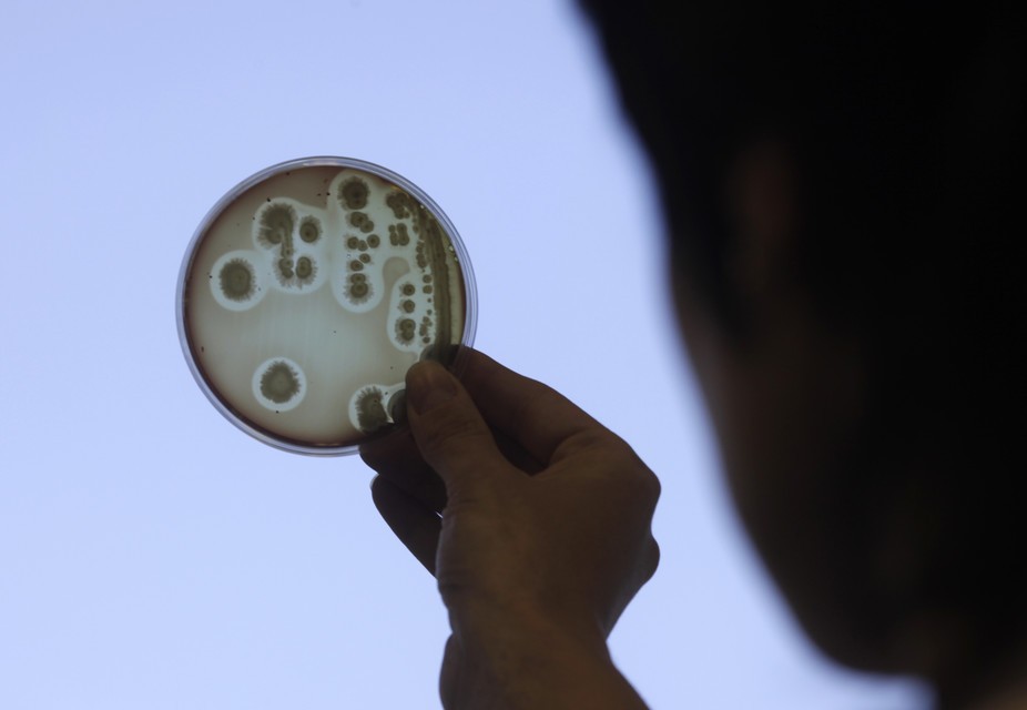
Stop the spread of superbugs,” “15 superbugs and other scary diseases” and “Superbug bacteria found in tested hotel rooms” are headlines we often read or hear about. But what do we mean when we say “bugs”?

The term is used to describe viruses, bacteria and parasites. While they can all make us sick, they do it in different ways. So what is the difference between these pathogens, and how dangerous are they?

Let’s start with viruses, the smallest of the three.


Electron microscope image of rabies virus (Rabdoviridae).
Sanofi Pasteur/Flickr, CC BY-NC-ND

Viruses – from the common cold to Ebola

Viruses have been around for a really, really long time. They predate us and could even be our oldest ancestors.

Viruses have helped build genomes of all species, including humans. Our genome is made up of 50 percent retroelements – the DNA from retroviruses. And viruses might have paved the way for several DNA replication enzymes, which are essential for a cell to divide and grow.

Viruses are capable of causing infections in humans and animals – and some viruses can even jump from one to the other.

Viruses have two phases of life. Outside a cell, they are nonliving and are called virion particles. Once inside a cell they use the cellular machinery to their advantage to replicate and multiply. Some scientists may argue that viruses are alive when inside a cell.

Some viruses, like the common cold, can make us sick, but don’t do lasting harm. But others are known to cause lethal disease in humans and animals. A pandemic strain of influenza can severely infect a large number of people in a very short time. There were an estimated 201, 200 respiratory deaths with an additional 83,300 cardiovascular deaths globally during the 2009 influenza (H1N1) pandemic.

While we are exposed to virus particles every day, we don’t always fall sick because the immune system can handle most of them. We get sick when we encounter a new virus for the first time or in sufficient quantity. This is why it is recommended to get a flu shot every year. The circulating strain of influenza may vary each year, and immunity from a previous infection or vaccine might not protect us in the event of exposure to a different strain.

The ability to spread quickly and replicate rapidly makes some of these viruses dreaded entries on the list of pathogens, to an extent that some are even considered as potential weapons of mass destruction. There are also viruses that kill slowly over time. A classic example is the rabies virus. It has a long incubation period (1-3 months) and is vaccine-preventable, but once the symptoms set in, the individual is almost certain to die.

Vaccines are the best way to protect ourselves from viruses. Vaccines prime the immune response, allowing our bodies to respond to an actual infection more efficiently. Vaccines have reduced the disease burden for several otherwise lethal viruses such as measles, rubella, influenza and smallpox. Beyond that, washing hands and covering noses while sneezing are practices that can keep some of these viruses at bay.


Immunohistochemical detection of Helicobacter histopatholgy. KGH via Wikimedia Commons, CC BY-SA

Bacteria – toxin-producing invaders

Some bacteria are good for you, offering protection against pathogens and aiding with digestion in the gut. But some aren’t so beneficial or benign.

Some are specialized to cause disease such as Staphylococcal infection (Staphylococcus aureus), botulism (Clostridium botulinum), gonorrhea (Neisseria gonorrhoeae), gastric ulcer (Helicobacter pylori), diphtheria (Corynebacterium diptheriae) and bubonic plague (Yersinia pestis).

They can produce toxins, invade cells or the bloodstream, or compete with the host for shared nutrients – all of which can lead to illness. The right course of treatment can depend on how the bacteria is causing illness.

Take botulism, for instance. People get it when they eat food contaminated with toxins or bacterial spores from C. botulinum. If a person ingests the toxin, he or she can develop symptoms within six to 36 hours. If the spore is ingested, it can take up to a week.

Supportive care is the primary therapeutic method, to prevent or relieve other possible complications and to maintain the health and breathing of the patient. Antibiotics treat infections by destroying the bacterium, but with botulism, the destruction of the bacterium can lead to the release of more toxins, causing severe illness. Doctors treat toxins by administering antitoxins or inducing vomiting.

Today, thanks to the misuse and overuse of antibiotics, resistant bacteria is on the rise, and as of 2013, there were about 480,000 new cases of multidrug-resistant tuberculosis (MDR-TB).

Cycling between different antibiotics can reduce the risk of resistance. Alternatives, such as bacteriophages (bacteria killing viruses) or enzymes that destroy the genome of resistant bacteria, are being developed. In fact, bacteriophages are widely used in Eastern Europe but haven’t been approved in North America.

There are vaccines available for some bacteria, like the DPT vaccine against Diphtheria, Bordetella pertussis and Clostridium tetani. And there are plenty of simple solutions to prevent bacteria from making us sick, such as proper hand washing, disinfection of surfaces, use of clean water and cooking to appropriate temperatures to eliminate bacteria.


Leishmania mexicana parasites.
Wellcome Images, CC BY-NC-ND

Parasites – benefiting at our expense

The third group in our trio of pathogens – parasites – have inspired many horror stories and many of us find them kind of gross.

Parasites are a diverse group of organisms that live in or on a host (like us) and benefit at the host’s expense. Parasites can be microscopic single cellular organisms called protozoa, or bigger organisms like worms or ticks. Protozoan parasites are actually more closely related to the cells in our body than to bacteria.

Parasites are everywhere, and they can play a complex and important role in ecosystems.

But parasites can also cause horrendous diseases, especially in the developing world. In many cases, infection with parasites goes hand in hand with bad sanitary conditions and poverty. Even though much progress has been made, malaria, which kills one child every 30 seconds with 90 percent of the cases in Africa, is still the most deadly disease caused by parasites. But it is by far not the only one.

Other parasitic diseases common in many – mostly tropical – parts of the world are Leishmaniasis, River Blindness and Elephantiasis.

Many parasites are transmitted by mosquitoes and other insects, and with the effects of climate change intensifying, many parasitic diseases are likely to move farther north.

Parasitic diseases are on the rise in developed countries, including the U.S. Chagas disease, for example, is caused by a single cellular parasite and cases are increasing in North America, possibly aided by climate change.

There are no vaccines available so far against any major parasitic diseases in humans, but there is plenty of research on that front. Luckily, there are many drugs available to combat parasites.

For instance, the 2015 Nobel Prize in Medicine was given to scientists who developed antiparasitic drugs (one drug, Ivermectin, treats worms; the other, Artemisinin, treats malaria).

These two drugs have helped whole countries to manage scourges caused by parasitic worms and malaria.

The latest success was in September 2015, when Mexico eliminated River Blindness, which is caused by Onchocerca volvulus, with the help of ivermectin donated by Merck.

Stay clean

Getting a harmful virus, bacterial infection or parasite disease isn’t good news. Fortunately we have effective treatments for some of them, and vaccines that can prevent us from getting sick as well, even if some of these bugs can evade the best medicines we have.

And keep in mind that even if these bugs can make us very, very sick, you still need to be exposed to them to become infected. While bigger strategies, like sanitation and infection control can keep us and others safe, so can simple strategies, like washing our hands, staying home when we are sick and covering our mouths when we cough or sneeze.

The ConversationArinjay Banerjee, PhD Student in Veterinary Microbiology, University of Saskatchewan; Jason Byron Perez, MSc Student, University of Saskatchewan, and Simona John von Freyend, Research Fellow, Malaria biochemistry, Monash University

This article was originally published on The Conversation. Read the original article.

Featured Image Credit: Ints Kalnins/Reuters

Now, Check Out: 

If we don’t own our genes, what protects study subjects in genetic research?

Leslie E. Wolf, Georgia State University; Erin Fuse Brown, Georgia State University, and Laura Beskow, Duke University

On February 25, the White House hosted a forum on the National Institute of Health’s Precision Medicine Initiative. This is an ambitious research study that aims to develop targeted drugs and treatments that would vary from individual to individual.

To reach the goal of eventually being able to make specific recommendations for patients based on their own combination of genes, environment and lifestyle, researchers plan to collect that kind of information from one million Americans. The study is so large so results can account for diversity among Americans with respect to factors such as ancestry, geography, and social and economic circumstances.

At the forum, President Obama remarked “I would like to think that if somebody does a test on me or my genes, that that’s mine.”

Lots of people would make that same assumption – it seems sensible that we would each “own” our genetic information. But the legal reality is quite different. And that could turn out to be a problem, because research projects like the Precision Medicine Initiative rely on research participants trusting that their information is protected once they agree to share it.

As scholars with expertise in research ethics, informed consent and health law, we’re conducting research to clarify how different laws apply to information used for genomic research. We’ll identify gaps in those protections and suggest changes that may be necessary.


President Obama discussing the Precision Medicine Initiative.
Carlos Barria/Reuters

Do you own your genes?

Contrary to President Obama’s expectations, the few U.S. courts that have considered research participants’ claims of ownership of their biological materials have rejected them.

  • John Moore’s doctor used his cells without his knowledge to develop and patent a cell line (cells that could continue to reproduce indefinitely for research). In 1990, the California Supreme Court held that Mr. Moore did not own the cells that had been removed from his body.
  • The Greenbergs and other families affected by Canavan disease, an inherited, degenerative and fatal brain disease in children, provided a University of Miami researcher with tissue and blood samples, medical information and money to develop a genetic test. The researcher patented the associated gene sequence, limiting families’ access to it without payment. In 2003, a federal court rejected the parents’ claims that they owned their genetic samples.
  • About 6,000 research participants responded to a letter sent by Dr. William Catalona, the developer of the prostate specific antigen test, and asked that their research samples stored at Washington University be transferred to Northwestern University, where Dr. Catalona had a new job. But a court determined that the research participants had no control over who held their specimens after collection.

The courts that have looked at the question have consistently decided that once we give our biological materials to researchers, the materials and the genetic information they contain belong to the researchers or, more specifically, the institutions that employ them.

A few states have adopted statutes concerning ownership of genes, but they may not alter court decisions. A Florida statute certainly did not make a difference in the Greenbergs’ case.

Short of ownership, what protections exist?

So you don’t own your genes. But there are other protections for participants in the Precision Medicine Initiative and other research projects.

The primary one comes from the Federal Common Rule. It applies to research conducted or funded by 18 federal departments and agencies. Many universities and other institutions apply the Common Rule to their research too. And research on drugs and devices that must be approved by the Food and Drug Administration (FDA) must comply with very similar rules.

Under the Common Rule, with some exceptions, research studies must be reviewed and approved by an Institutional Review Board (IRB): a committee within the university or hospital, for instance, that scrutinizes proposed experiments involving human subjects. In approving a study, the IRB must evaluate, among other things, the adequacy of the consent process and confidentiality protections, whether risks are minimized and are reasonable in relation to the benefits, and whether the selection of subjects is equitable. The IRB provides a check on what researchers can do.

Once the Institutional Review Board approves a study, researchers can start recruiting people to participate. This is where another protection comes in – consent.


Study subjects should understand the potential risks and benefits of participating.
Form image via www.shutterstock.com.

The researchers must disclose the research’s purpose, procedures and any risks and benefits of participating. In a study like the Precision Medicine Initiative, the primary risks are informational, not physical. For example, if an insurer learned that a research participant had a gene that increases the risk of Alzheimer’s, it might refuse long-term care coverage.

Based on the risks and benefits (if any) discussed in the consent form, participants can decide whether they want to take part. They may decline to participate if they do not trust the researchers or do not want to share their information.

In some circumstances, the Common Rule doesn’t require participant consent. These exceptions are allowed when the study poses little risk to the participant, often because the information cannot be connected to the individual.

In recent years, these exceptions have been called into question as researchers have repeatedly demonstrated that it is possible to identify people whose information has been used in research, but were thought to be unidentifiable. However, such reidentification requires significant effort and technical skills, and, alone, is unlikely to result in harm to participants. Thus, it is not clear that we should forego the benefits of research conducted under these exceptions because of the theoretical threat to confidentiality.

Beyond these exceptions, some research – such as Facebook’s 2014 study that manipulated some 700,000 users’ newsfeeds to determine the effect of negative or positive words on their emotions – falls outside the Common Rule altogether.

In general, research that is not federally conducted or funded or subject to FDA regulations is not governed by federal research protections. Some states have adopted laws that apply similar protections to research not subject to either the Common Rule or the FDA regulations, but those laws vary considerably from state to state.

Additional protections for research participants

The Health Insurance Portability and Accountability Act’s (HIPAA) privacy rule provides a national standard for protecting the use and disclosure of identifiable health information. The corresponding security rule establishes standards for securing electronic health records which could include results of genetic research.

In addition, the Genetic Information Nondiscrimination Act (GINA) prohibits use of genetic information to discriminate against asymptomatic individuals in employment and health insurance decisions. Although it has recognized gaps, GINA provides some protections against discrimination, should genetic information from a research study be disclosed.

As with the Common Rule, state medical privacy and antidiscrimination laws may supplement these federal protections. Thus, the protections afforded to participants may depend greatly on where they live. Moreover, Institutional Review Boards may be unfamiliar with the myriad laws that could combine to protect research participants and their possible gaps.

Beyond these legal requirements, the Precision Medicine Initiative may provide participants additional controls over their data on a voluntary basis. For example, participants could reevaluate their preferences for how their data are shared or used, withdraw their consent for future use of their data at any time and control the types of communications they receive about their information.

While these types of protections may fall short of full legal ownership rights over your genetic information, they do go beyond current legal requirements and may be the types of controls to which President Obama was alluding.


The samples have been collected… now what happens?
Geir Mogen, NTNU, CC BY-NC

What is needed?

We think it is essential for all those involved in research – IRBs, researchers and study participants – to understand what protections are available and what their limitations are.

That’s why we’ve undertaken a comprehensive analysis of federal and state laws that combine to form what we call the “web of protections.” We want to be able to describe how the laws work together, to identify gaps, and to suggest ways to improve those protections, as well as how all this should be described to prospective research participants.

To the extent that the current laws fall short of the types of protections and controls expected by participants in research studies like the Precision Medicine Initiative, we may be able to propose ways that the laws can be updated or supplemented to address concerns like President Obama’s. In this way, we can maintain the public trust on which this research relies.

The ConversationLeslie E. Wolf, Professor of Law and Director, Center for Law Health and Society, College of Law, Georgia State University; Erin Fuse Brown, Assistant Professor of Law, Georgia State University, and Laura Beskow, Director of the Program for Empirical Bioethics, Associate Professor of Medicine, Duke University

This article was originally published on The Conversation. Read the original article.

Featured Image Credit: gemhq, CC BY

Now, Check Out:

Here’s Why Geysers On Saturn’s Moon Have Nonstop Eruptions

The Cassini spacecraft has observed geysers erupting on Saturn’s moon Enceladus since 2005, but the process that drives and sustains these seemingly endless eruptions has remained a mystery.

Now scientists have pinpointed the mechanism by which cyclical tidal stresses exerted by Saturn can drive those long-lived eruptions.

“On Earth, eruptions don’t tend to continue for long,” says Edwin Kite, assistant professor of geophysical sciences at the University of Chicago. “When you see eruptions that continue for a long time, they’ll be localized into a few pipe-like eruptions with wide spacing between them.”

But Enceladus, which probably has an ocean underlying its icy surface, has somehow managed to sprout multiple fissures along its south pole. These “tiger stripes” have been erupting vapor and tiny frost particles continuously along their entire length for decades and probably much longer.

casini-eruptions
Jets of icy particles burst from Saturn’s moon Enceladus in this sequence of four images taken from the Cassini spacecraft on Nov. 27, 2005. The geysers of Enceladus continue to erupt today. (Credit: NASA/JPL/Space Science Institute)

A LEADING CANDIDATE FOR LIFE

“It’s a puzzle to explain why the fissure system doesn’t clog up with its own frost,” Kite says. “And it’s a puzzle to explain why the energy removed from the water table by evaporative cooling doesn’t just ice things over.”

What’s needed is an energy source to balance the evaporative cooling. “We think the energy source is a new mechanism of tidal dissipation that had not been previously considered,” says Kite, coauthor with Allan Rubin of Princeton University of a new study published in the Proceedings of the National Academy of Sciences.

“I was very happy to see this new work by Kite and Rubin that brings to the fore a process that had escaped notice: the pumping of water in and out of the deep fractures of the south polar ice shell by tidal action,” says Carolyn Porco, head of Cassini’s imaging science team and a leading scientist in the study of Enceladus.

Enceladus, which Kite calls “an opportunity for the best astrobiology experiment in the solar system,” serves as a leading candidate for extraterrestrial life. Cassini data have strongly indicated that the cryovolcanic plumes of Enceladus probably originate in a biomolecule-friendly oceanic environment.

Cryovolcanism also may have shaped the surface of Europa, one of Jupiter’s moons. “Europa’s surface has many similarities to Enceladus’s surface, and I hope this model will be useful for Europa as well,” Kite says.

One of the problems that attracted Kite and Rubin was the anomalous tidal response of the Enceladus eruptions. The eruptions reach their peak approximately five hours later than expected—even when taking into account the 40 minutes needed for the erupted particles to reach the altitude at which Cassini can detect them. Scientists had previously suggested reasons for the lag, which included a delay in the eruptions as well as a squishy, slowly responding ice shell.

“The new proposal is really a way to get a delay in the eruptions. You really don’t need to propose any terribly squishy ice shell to do it,” Porco says.

Kite and Rubin also wanted to know why Enceladus maintains a base level of cryovolcanic activity, even when at that point in its orbit where the fissures should clamp shut and curtail the eruptions. Other key questions: Why does the volcanic system generate five gigawatts of power instead of a lot more or a lot less? Why don’t the eruptions frost over or freeze over?

PLUMBING ANSWERS THE QUESTIONS

The new computer model of the Enceladus plumbing system seems to answer them all. Their model consists of a series of nearly parallel, vertical slots that reach from the surface down to the water below. They applied Saturn’s tidal stresses to their model on a desktop computer and watched what happened.

“The only tricky part quantitatively is calculating the elastic interactions between the different slots and the varying water level within each slot as a response to the tidal stress,” Kite says. The width of the slots affects how quickly they can respond to the tidal forces. With wide slots, the eruptions respond quickly to tidal forcing. With narrow slots, the eruptions occur eight hours after the tidal forces reach their peak.

“In between there’s a sweet spot,” Kite says, where tidal forces turn water motion into heat, generating enough power to produce eruptions that match the observed five-hour lag. Porco called it “the best thing in my mind about this new work.”

Tidal pumping heats the water and the ice shell via turbulence. Kite and Rubin have proposed that new Cassini data can test this idea by revealing whether the ice shell in the south polar region is warm.

“If the new mechanism is a major contributor to the heat coming from the fractures, then the south polar ice in between the fractures may, in fact, be cold,” Porco says. Results from last year’s Enceladus flyby need to be analyzed to make that determination.

Kite and Douglas MacAyeal, professor of geophysical sciences at the University of Chicago are interested in studying an Earth analogue to the Enceladus geysers. A crack has formed across a section of the Ross Ice Shelf in Antarctica, partially breaking it away from the continent.

“In that crack you have strong tidal flow, so it would be interesting to see what a real ice sheet does in an environment that’s analogous in terms of the amplitude of the stresses and the temperatures of the ice,” Kite says.

NASA funded the work; the study is published in the Proceedings of the National Academy of Sciences.

Source: Republished from Futurity.org as a derivative work under the Attribution 4.0 International license. Original article posted to Futurity by .

Featured Photo Credit: NASA/JPL/Space Science Institute

Now, Check Out:

What if the FBI tried to crack an Android phone? We attacked one to find out

By William Enck, North Carolina State University and Adwait Nadkarni, North Carolina State University.

The Justice Department has managed to unlock an iPhone 5c used by the gunman Syed Rizwan Farook, who with his wife killed 14 people in San Bernardino, California, last December. The high-profile case has pitted federal law enforcement agencies against Apple, which fought a legal order to work around its passcode security feature to give law enforcement access to the phone’s data. The FBI said it relied on a third party to crack the phone’s encrypted data, raising questions about iPhone security and whether federal agencies should disclose their method.

But what if the device had been running Android? Would the same technical and legal drama have played out?

We are Android users and researchers, and the first thing we did when the FBI-Apple dispute hit popular media was read Android’s Full Disk Encryption documentation.

We attempted to replicate what the FBI had wanted to do on an Android phone and found some useful results. Beyond the fact the Android ecosystem involves more companies, we discovered some technical differences, including a way to remotely update and therefore unlock encryption keys, something the FBI was not able to do for the iPhone 5c on its own.

The easy ways in

Data encryption on smartphones involves a key that the phone creates by combining 1) a user’s unlock code, if any (often a four- to six-digit passcode), and 2) a long, complicated number specific to the individual device being used. Attackers can try to crack either the key directly – which is very hard – or combinations of the passcode and device-specific number, which is hidden and roughly equally difficult to guess.

Decoding this strong encryption can be very difficult. But sometimes getting access to encrypted data from a phone doesn’t involve any code-breaking at all. Here’s how:

  • A custom app could be installed on a target phone to extract information. In March 2011, Google remotely installed a program that cleaned up phones infected by malicious software. It is unclear if Android still allows this.
  • Many applications use Android’s Backup API. The information that is backed up, and thereby accessible from the backup site directly, depends on which applications are installed on the phone.
  • If the target data are stored on a removable SD card, it may be unencrypted. Only the most recent versions of Android allow the user to encrypt an entire removable SD card; not all apps encrypt data stored on an SD card.
  • Some phones have fingerprint readers, which can be unlocked with an image of the phone owner’s fingerprint.
  • Some people have modified their phones’ operating systems to give them “root” privileges – access to the device’s data beyond what is allowed during normal operations – and potentially weakening security.

But if these options are not available, code-breaking is the remaining way in. In what is called a “brute force” attack, a phone can be unlocked by trying every possible encryption key (i.e., all character combinations possible) until the right one is reached and the device (or data) unlocks.

Starting the attack


A very abstract representation of the derivation of the encryption keys on Android.
William Enck and Adwait Nadkarni, CC BY-ND

There are two types of brute-force attacks: offline and online. In some ways an offline attack is easier – by copying the data off the device and onto a more powerful computer, specialized software and other techniques can be used to try all different passcode combinations.

But offline attacks can also be much harder, because they require either trying every single possible encryption key, or figuring out the user’s passcode and the device-specific key (the unique ID on Apple, and the hardware-bound key on newer versions of Android).

To try every potential solution to a fairly standard 128-bit AES key means trying all 100 undecillion (1038) potential solutions – enough to take a supercomputer more than a billion billion years.

Guessing the passcode could be relatively quick: for a six-digit PIN with only numbers, that’s just a million options. If letters and special symbols like “$” and “#” are allowed, there would be more options, but still only in the hundreds of billions. However, guessing the device-specific key would likely be just as hard as guessing the encryption key.

Considering an online attack

That leaves the online attack, which happens directly on the phone. With the device-specific key readily available to the operating system, this reduces the task to the much smaller burden of trying only all potential passcodes.

However, the phone itself can be configured to resist online attacks. For example, the phone can insert a time delay between a failed passcode guess and allowing another attempt, or even delete the data after a certain number of failed attempts.

Apple’s iOS has both of these capabilities, automatically introducing increasingly long delays after each failure, and, at a user’s option, wiping the device after 10 passcode failures.

Attacking an Android phone

What happens when one tries to crack into a locked Android phone? Different manufacturers set up their Android devices differently; Nexus phones run Google’s standard Android configuration. We used a Nexus 4 device running stock Android 5.1.1 and full disk encryption enabled.


Android adds 30-second delays after every five failed attempts; snapshot of the 40th attempt.
William Enck and Adwait Nadkarni, CC BY-ND

We started with a phone that was already running but had a locked screen. Android allows PINs, passwords and pattern-based locking, in which a user must connect a series of dots in the correct sequence to unlock the phone; we conducted this test with each type. We had manually assigned the actual passcode on the phone, but our unlocking attempts were randomly generated.

After five failed passcode attempts, Android imposed a 30-second delay before allowing another try. Unlike the iPhone, the delays did not get longer with subsequent failures; over 40 attempts, we encountered only a 30-second delay after every five failures. The phone kept count of how many successive attempts had failed, but did wipe the data. (Android phones from other manufacturers may insert increasing delays similar to iOS.)

These delays impose a significant time penalty on an attacker. Brute-forcing a six-digit PIN (one million combinations) could incur a worst-case delay of just more than 69 days. If the passcode were six characters, even using only lowercase letters, the worst-case delay would be more than 58 years.

When we repeated the attack on a phone that had been turned off and was just starting up, we were asked to reboot the device after 10 failed attempts. After 20 failed attempts and two reboots, Android started a countdown of the failed attempts that would trigger a device wipe. We continued our attack, and at the 30th attempt – as warned on the screen and in the Android documentation – the device performed a “factory reset,” wiping all user data.


Just one attempt remaining before the device wipes its data.
William Enck and Adwait Nadkarni, CC BY-ND

In contrast to offline attacks, there is a difference between Android and iOS for online brute force attacks. In iOS, both the lock screen and boot process can wipe the user data after a fixed number of failed attempts, but only if the user explicitly enables this. In Android, the boot process always wipes the user data after a fixed number of failed attempts. However, our Nexus 4 device did not allow us to set a limit for lock screen failures. That said, both Android and iOS have options for remote management, which, if enabled, can wipe data after a certain number of failed attempts.

Using special tools

The iPhone 5c in the San Bernardino case is owned by the employer of one of the shooters, and has mobile device management (MDM) software installed that lets the company track it and perform other functions on the phone by remote control. Such an MDM app is usually installed as a “Device Administrator” application on an Android phone, and set up using the “Apple Configurator” tool for iOS.


Our test MDM successfully resets the password. Then, the scrypt key derivation function (KDF) is used to generate the new key encryption key (KEK).
William Enck and Adwait Nadkarni, CC BY-ND

We built our own MDM application for our Android phone, and verified that the passcode can be reset without the user’s explicit consent; this also updated the phone’s encryption keys. We could then use the new passcode to unlock the phone from the lock screen and at boot time. (For this attack to work remotely, the phone must be on and have Internet connectivity, and the MDM application must already be programmed to reset the passcode on command from a remote MDM server.)

Figuring out where to get additional help

If an attacker needed help from a phone manufacturer or software company, Android presents a more diverse landscape.

Generally, operating system software is signed with a digital code that proves it is genuine, and which the phone requires before actually installing it. Only the company with the correct digital code can create an update to the operating system software – which might include a “back door” or other entry point for an attacker who had secured the company’s assistance. For any iPhone, that’s Apple. But many companies build and sell Android phones.

Google, the primary developer of the Android operating system, signs the updates for its flagship Nexus devices. Samsung signs for its devices. Cellular carriers (such as AT&T or Verizon) may also sign. And many users install a custom version of Android (such as Cyanogenmod). The company or companies that sign the software would be the ones the FBI needed to persuade – or compel – to write software allowing a way in.

Comparing iOS and Android

Overall, devices running the most recent versions of iOS and Android are comparably protected against offline attacks, when configured correctly by both the phone manufacturer and the end user. Older versions may be more vulnerable; one system could be cracked in less than 10 seconds. Additionally, configuration and software flaws by phone manufacturers may also compromise security of both Android and iOS devices.

But we found differences for online attacks, based on user and remote management configuration: Android has a more secure default for online attacks at start-up, but our Nexus 4 did not allow the user to set a maximum number of failed attempts from the lock screen (other devices may vary). Devices running iOS have both of these capabilities, but a user must enable them manually in advance.

Android security may also be weakened by remote control software, depending on the software used. Though the FBI was unable to gain access to the iPhone 5c by resetting the password this way, we were successful with a similar attack on our Android device.

The ConversationWilliam Enck, Assistant Professor of Computer Science, North Carolina State University and Adwait Nadkarni, Ph.D. Student of Computer Science, North Carolina State University

This article was originally published on The Conversation. Read the original article.

Now, Check Out:

Resurrected Drug Surprises Researchers with Powerful, Broad-Spectrum Antiviral Potential

Viruses have proven to be wily foes. Attempts to fend off viruses that cause the common cold or flu have failed, and new viral outbreaks such as dengue, Ebola, or Zika continue to elude drugs.

Given these challenges, scientists are tackling the problem from a different angle. They want to boost the human body’s ability to resist the virus rather than taking on the virus directly.

Now, it appears the approach has paid off with a drug that—in cells in a lab dish at least—helps fight two disease-causing viruses and potentially many more.

The way the drug works suggests that it could be broadly effective against viruses that use RNA rather than DNA as their genetic material, says Chaitan Khosla, professor of chemistry and of chemical engineering at Stanford University.

ONE DRUG, MULTIPLE BUGS

“Most of the really nasty viruses use RNA,” including Ebola, dengue, Zika, and Venezuelan equine encephalitis virus (VEEV), a mosquito-borne virus that infects horses but can also kill people.

Khosla cautions that at this stage the drug has only been shown to be effective in a lab dish and on certain viruses, but researchers plan to test the strategy in animals next to learn whether it is safe and to understand which viral diseases it is most effective against.

The project came about when Jeffrey Glenn, associate professor of medicine and of microbiology and immunology, founded the ViRX@Stanford center through a grant from the National Institute of Allergy and Infectious Diseases in collaboration with Stanford ChEM-H, which Khosla directs. The center’s goal is to develop antiviral strategies targeting human cells rather than the virus.

Scientists typically take a “one drug, one bug” approach to fighting viruses. The center, however, has a goal of “one drug, multiple bugs.”

The team had known about a drug being developed by GlaxoSmithKline that appeared to work along these lines, helping human cells fight viruses. However, after a few initial publications the drug got shelved. Researchers thought that with the help of collaborations formed through the new center, it might be possible to understand the drug’s mechanism and possibly improve upon it, resurrecting the drug from the shelves and delivering it to patients.

Chemistry graduate student Richard Deans started testing that drug on human cells in a lab dish and found that it enabled the cells to fight off viruses that cause either dengue or VEEV, both of which normally kill the cells. These viruses were chosen because they represent a serious threat to human health, and also represent two different classes of RNA viruses and would test the drug’s breadth, according to Jan Carette, assistant professor of microbiology and immunology and an author on the paper.

Although the drug was effective at fighting the viruses, Deans found that over time the drug also caused the human cells to stop dividing.

As a first step to improving on the drug, Deans needed to figure out how it worked. For that, he turned to Michael Bassik, assistant professor of genetics and a senior author on the paper.

DRUG RESPONSE

Bassik, who is also member of Stanford Bio-X and ChEM-H, had developed a powerful new way of screening every gene in a cell to identify which proteins those genes produce to carry out a particular behavior, like responding to a drug.

From this screen, the team learned that the drug interferes with a protein that is crucial for making the individual building blocks of RNA, the genetic code for the virus. Without RNA the virus can’t make more of itself, which explains why the drug was so effective.

However, because of the way the screen was designed, it also revealed two important additional details that the team wouldn’t have otherwise known: why the drug doesn’t work perfectly and why it causes cells to stop dividing. That information gave the team a way of reducing the drug’s side effects and also suggested a way of making it more effective.

“The genome-wide screen carried out in the Bassik lab was really powerful, because it gave us insights into future research strategies,” says Deans, who was lead author of the paper, that is published in Nature Chemical Biology. “I think going forward his strategy will be much more heavily used.”

Cells also need RNA, and can get RNA building blocks in two ways—by making them or by importing them from the bloodstream. The drug blocked the cell’s ability to make the RNA building blocks but left intact the cell’s ability to import them. Without disrupting both pathways, some RNA precursors made it into the cell and were available to the virus.

The team is now testing their drug along with another one that is known to block the import pathway to see if the combination is more effective than one drug alone, and to be sure human cells aren’t damaged by the absence of RNA building blocks.

KNOW THE PROBLEM, FIND A SOLUTION

Knowing how the drug works also explains why it can cause the body’s normal, healthy cells to stop dividing. The same building blocks needed for RNA synthesis are also needed to make DNA, the cell’s genetic code that it replicates with each division to carry out business as usual. When a cell runs out of DNA building blocks, it can no longer divide.

Knowing the problem, the team could devise a solution. They fed the cells a slightly different building block that can only be used to generate DNA, not RNA. With that added to the mix, the cells successfully fought off both dengue and VEEV and were able to keep dividing normally. This knowledge could help make the drug less toxic in animals and eventually people.

The researchers plan to test the drug combination against many different RNA viruses to learn which it fights most effectively. If the drug combination is successful in animals, they hope it might become among the first broad antiviral strategies for human disease.

The National Science Foundation Graduate Research Fellowship, a Burt and Deedee McMurtry Stanford Graduate Fellowship, the National Institutes of Health, a Director’s New Innovator Award Program,  and a seed grant from Stanford ChEM-H funded the work.

Source: Republished from Futurity.org as a derivative work under the Attribution 4.0 International license. Original article posted to Futurity by .

Featured Image Credit:  Leonel Cunha/Flickr, CC BY-2.0

Now, Check Out:

What fish mouths teach us about engineering clog-free filters [Video]

S. Laurie Sanderson, College of William & Mary

Filter-feeding fish accomplish a feat that human technologies cannot: species including goldfish, menhaden and basking sharks filter tiny algal cells or shrimp-like prey from huge volumes of water without clogging their oral filters.

Since fish have been filtering particles for more than 150 million years longer than human beings, we suspected fish may have evolved filter designs that use unknown processes to remain unclogged. So we decided to investigate.

Our research, recently published in Nature Communications, combines approaches from biomechanics, medicine and ecology to explore how these fish retain and transport prey inside their mouths. Our goal is to provide ideas and data that could improve aquaculture, conservation and industrial filtration.

Crossflow filtration works for fish and industry

Until 15 years ago, we thought that most filter-feeding fish used oral structures called gill rakers in the same way that we use coffee filters or spaghetti strainers. These so-called dead-end sieves force water to pass straight through the pores of the mesh. But dead-end sieves always clog as particles accumulate over time to cover the filter surface.

The water flows right through a colander and leaves the spaghetti trapped on the mesh, but a fish needs to move the food from the gill raker filter to the back of its mouth for swallowing. Dead-end sieves would cause problems for fish, since their gill rakers would clog and fish don’t have a tongue to move food particles off the gill rakers. So we knew they must be using some other filtering technique.

By putting a biomedical endoscope inside the mouths of feeding fish, colleagues and I discovered in 2001 that several common fish species use crossflow filtration instead of trapping particles directly on a dead-end sieve.

During crossflow filtration, small secondary streams of fluid pass through each filter pore – perpendicular to the filter surface, like in dead-end filtration. But the main stream of fluid – the “crossflow” – is directed to travel across (parallel to) the filter surface, lifting particles off the filter and preventing the pores from clogging with particles.

A tilapia illustrating the current model of crossflow filtration, from Sanderson et al., doi: 10.1038/ncomms11092. The mainstream flow (MF) enters from the right and passes across the gill rakers (GR) that are attached to the branchial arches (BA). The mainstream flow carries concentrated particles to the back of the mouth for swallowing. The smaller secondary flows (the filtrate, Fi) pass through the pores of the gill raker filter. Virginia Greene, virginiagreeneillustration.com, CC BY-NC-ND

Through the endoscope, we could see that the main flow of water heading toward the back of the mouth was transporting concentrated particles parallel to the gill raker filter. Less forceful streams of particle-free water exited between the gill rakers. All of these fluid dynamics are caused by the interaction of the water with the physical structures in the fish’s mouth.

We hadn’t expected to see crossflow filtration in fish, though this mechanism had been independently developed by industry a few decades earlier. Crossflow filtration avoids clogging and is often used to filter wastewater, pharmaceuticals, dairy foods and beverages such as beer and fruit juices.

Unfortunately, even industrial crossflow filters still clog eventually. Over time, as water exits through the filter pores, it deposits some particles on the filter. The filters must then be backflushed or cleaned with chemicals, causing a major operating expense.

So we turned again to fish, to see whether millions of years of evolution might have come up with unique crossflow filter designs.

Biomimetic designs from fish mouths

We started our study by examining basic structures inside fish mouths, familiar to fishermen and aquarium hobbyists. Fish gill rakers – the “feeding filters” – are attached to the branchial arches. These arches are bone or cartilage “ribs” inside the mouth that also support the bright red gills for gas exchange. The arches are typically positioned one after another from the front of the mouth back toward the esophagus, where food is swallowed. Scientists hadn’t previously considered the effects these branchial arches could have on patterns of water flow.

For our latest research, we made our own filters by using computer-aided design (CAD) software and 3D printing to create cone-shaped plastic models of fish mouths. We covered the branchial arch “ribs” with a fine nylon mesh.

We based our physical models on paddlefish and basking sharks because their branchial arches form a series of tall ribs that are separated by deep grooves. In our models, each rib served as a backward-facing step that interacted with the crossflow of water traveling over the step.

Almost anywhere that water flows over a backward-facing step, a vortex is created automatically. For this reason, the closely-spaced tall ribs (“d-type ribs”) in these fish mouths aren’t often used by engineers because of the disruptive vortices that form continuously in the grooves between the ribs.

We designed many models with different versions of these backward-facing steps to test the effects of varying characteristics like height and distance between the steps. Interestingly, designs for some microfluidics devices that are used in labs for cell sorting have similar rib-like structures.

Both paddlefish and basking sharks are ram filter feeders that swim forward with a completely open mouth to capture prey. To simulate this kind of feeding, my three undergraduate student coauthors, Erin Roberts, Jillian Lineburg and Hannah Brooks, and I conducted experiments in a flow tank. We submerged our stationary models in a constant stream of water inside the tank. The models “fed” on particles as we adjusted the speed of the water in the flow tank and added particles of different sizes, shapes and densities to the water.

A paddlefish illustrating the new vortical cross-step filtration model, from Sanderson et al., doi: 10.1038/ncomms11092. The mainstream flow (MF) enters from the right and interacts with the series of backward-facing steps that are formed by the branchial arches (BA), causing vortical flow (Vo). The vortex interacts with the gill rakers (GR) to concentrate particles for transport towards the back of the mouth to be swallowed. Virginia Greene, virginiagreeneillustration.com, CC BY-NC-ND

Unique vortical cross-step filtration in fish

Like the spinning of a mini-tornado, water passed over the backward-facing steps inside our models and formed a distinct vortex in the groove between each pair of ribs. We designed accessory structures to control the movement of the vortices by creating regions of the model where the flow couldn’t escape easily. High shear rates around the vortices scoured particles off the mesh, preventing clogging.

Green dye helps visualize the vortices generated in model paddlefish and basking shark mouths. S. Laurie Sanderson, CC BY-ND

We manipulated the vortices to carry particles to the floor of the models, showing that fish could be using this highly adaptable filtration system like a “hydrodynamic tongue” to move particles inside their mouths.

We manipulated the vortices in our models to transport concentrated particles along the vortex axis, downstream from each backward-facing step. The vortices lifted particles from the mesh and carried them toward the floor of the model.

Small preserved paddlefish from an aquaculture company, placed in the flow tank in filter-feeding position, also formed vortices that concentrated particles inside the mouth. This suggests that we’ve correctly identified and modeled structures that are important for generating vortices inside real fish mouths.

This new filtration method, which we term “vortical cross-step filtration,” is effective even when the mesh is damaged or missing from a large portion of the models. Just like fish can continue to feed even when their gill rakers are still growing or are torn, our models can capture particles even when there are large holes in the mesh.

Although we’d identified vortices as a potential mechanism for fish filtration as early as 2001, data on particle capture by vortical flow in fish mouths haven’t been published previously.

Rhodamine dye traces the path of a vortex that forms downstream from a backward-facing step. The step mimics a branchial arch inside a fish’s mouth.

The future of cross-step filtration

Our biomimetic models of paddlefish and basking shark mouths use novel arrangements of engineering structures that harness vortical flow to retain and transport tiny food particles. Cross-step filtration could also apply to filter-feeding ducks, baleen whales and the gill rakers of filter-feeding fish such as manta rays.

Understanding these vortices in fish opens new research directions for engineering improved filters with less clogging, as well as the rapid separation of cells for biomedical tests.

The ConversationS. Laurie Sanderson, Professor of Biology, College of William & Mary

This article was originally published on The Conversation. Read the original article.

Featured Photo Credit: Rob Holm / USFWS, CC BY

Now, Check Out:

Is global warming causing marine diseases to spread?

Charlotte Eve Davies, Universidad Nacional Autónoma de México (UNAM)

Global climate change is altering the world’s oceans in many ways. Some impacts have received wide coverage, such as shrinking Arctic sea ice, rising sea levels and ocean warming. However, as the oceans warm, marine scientists are observing other forms of damage.

My research focuses on diseases in marine ecosystems. Humans, animals and plants are all susceptible to diseases caused by bacteria, viruses, parasites and fungi. Marine diseases, however, are an emerging field.

Infectious agents have the potential to alter ocean life in many ways. Some threaten our food security by attacking important commercial species, such as salmon. Others, such as bacteria in oysters, may directly harm human health. Still others damage valuable marine ecosystems – most notably coral reefs.

To anticipate these potential problems, we need a better understanding of marine diseases and how climate change affects their emergence and spread.

Warming waters promote marine diseases

Recent studies show that for some marine species diseases are spreading and increasing. Climate change may also promote the spread of infectious agents in oceans. Notably, warming water temperatures can expand these agents’ ranges and introduce diseases to areas where they were previously unknown.

Many diseases of marine species are secondary opportunist infections that take advantage when a host organism is stressed by other conditions, such as changes in pH, salinity or temperature. A bacterium that is dormant (and therefore noninfective) at a certain temperature may thrive at a slightly higher temperature.

One well-documented example is the emergence of epizootic shell disease (ESD) in American lobsters. This disease, thought to be caused by bacteria, is characterized by lesions that penetrate inward from a lobster’s shell surface towards the inner flesh, making infected lobsters unmarketable. ESD can also kill lobsters by making it difficult for them to shed their shells in order to grow.

An American lobster with epizootic shell disease (ESD). para_sight/flickr

In the 1990s, following almost a decade of above-normal summer temperatures, ESD affected so many lobsters that the Atlantic States Marine Fisheries Commission declared that the Southern New England fishery (Connecticut, Massachusetts, New York and Rhode Island) was in collapse and recommended closing it.
Fishery models that incorporated shell disease offered convincing evidence that ESD was a major factor in the decline of the stock. This episode underscores the importance of considering marine diseases in stock assessments and fishery management.

Now there are concerns that ESD will continue to spread north to Maine’s US$465.9 million lobster fishery. In 2015 the Gulf of Maine showed record high abundances of lobster, making it one of the most productive fisheries in the world.

However, sea surface temperatures in the Gulf of Maine have increased faster than 99 percent of the global ocean over the past decade, warming three times faster than the global average. Since temperature is a primary factor in the spread of this disease, observers fear that it could have devastating effects on Maine’s lobster fishery.

There is also a risk that ESD could spread from American lobsters to other fisheries. Seafood wholesalers have imported live American lobsters into Europe for decades, which can result in their escape into the wild. Last summer the United Kingdom’s Marine Management Organization warned U.K. fishermen that because the European lobster shares similar habitats, food sources and diseases with the American lobster, ESD could spread between the species.

As a doctoral student at Swansea University, U.K., I collaborated with the New England Aquarium in Boston, Massachusetts to investigate this possibility. While we found that European lobsters were more likely to develop shell disease when reared in the presence of American lobsters, on the positive side, they don’t seem to get the same shell disease as American lobsters.

This means that European lobsters may be better equipped to deal with outbreaks of ESD. But with sea surface temperatures in U.K. coastal waters rising since the 1980s by around 0.2-0.9 degrees Celsius per decade, it is important to monitor U.K. waters for this disease.

European lobsters with mild, none and severe shell disease. Andrew Rowley/Swansea University

Tropical disease

Now I am now studying the Panuliris argus_1 virus (PaV1) in the Caribbean spiny lobster, where the picture is more dire. Discovered around 2000, this virus is present from the Florida Keys to Venezuela. It can infect up to 60 percent of lobsters in some areas. Laboratory studies indicate that lobsters held in high-temperature seawater and exposed to PaV1 develop active and more intense infections much more quickly than those held at lower temperatures.

Studies from 1982 to 2012 show that waters in the Caribbean are warming, with the most significant temperature increase occurring over the past 15 years – approximately the period when PaV1 appeared. If PaV1 continues to spread, it could have significant effects on the health of Caribbean reefs as a whole, as well as on the valuable Caribbean lobster fishery.

Monitoring more diseases

Many other species are also showing increasing effects from marine diseases. The frequency of coral diseases has increased significantly over the last 10 years, causing widespread mortality among reef-building coral, which are home to more than 25 percent of all marine fish species.

In the Pacific, more than 20 species of sea stars were devastated by a wasting disease that ranged from Mexico all the way up to Alaska in 2013 and 2014. Research suggests that 90 percent of some populations were wiped out, and some adult populations have been reduced to a quarter of pre-outbreak numbers.

Scientists believe the cause is a virus which becomes more active in warmer conditions. In both field surveys and laboratory experiments, starfish were found to react faster to the disease in warmer water than in cooler temperatures.

Starfish on the shore at Umpqua Lighthouse State Park – Winchester Bay, Oregon. skipplitt/flickr

As the oceans continue to warm, it is crucial to understand how our actions are affecting marine life. Some species will not be able to withstand the increase in temperature. The most recent U.S. National Climate Change Assessment projects that outbreaks of marine diseases are likely to increase in frequency and severity as waters warm under climate change. Researchers are working around the world to determine whether and how species will survive disease events in our increasingly altered oceans.

The ConversationCharlotte Eve Davies, Postdoctoral Researcher at the Institute of Marine Sciences and Limnology, Universidad Nacional Autónoma de México (UNAM)

This article was originally published on The Conversation. Read the original article.

Featured Photo Credit: James St. John/Flickr, CC BY

Now, Check Out:

Breakthrough ‘Fiber Islands’ 100 Times More Effective for Treating Brain Diseases

Biomedical scientists have figured out how to convert adult tissue-derived stem cells into human neurons on 3D “scaffolds,” or tiny islands of fibers.

The technology could someday help treat people with Parkinson’s disease and other devastating brain-related conditions, researchers say.

For a new study, scientists injected the scaffolds, loaded with beneficial neurons to replace diseased cells in mouse brains.

“If you can transplant cells in a way that mimics how these cells are already configured in the brain, then you’re one step closer to getting the brain to communicate with the cells that you’re now transplanting,” says Prabhas V. Moghe, professor of biomedical engineering and chemical and biochemical engineering at Rutgers University. “In this work, we’ve done that by providing cues for neurons to rapidly network in 3D.”

Neurons, or nerve cells, are critical for human health and functioning. Human brains have about 100 billion neurons, which serve as messengers that transmit signals from the body to the brain and vice versa.

The new 3D scaffold consists of tiny polymer fibers. Hundreds of neurons attach to the fibers and branch out, sending their signals. Scaffolds are about 100 micrometers wide—roughly the width of a human hair.

“We take a whole bunch of these islands and then we inject them into the brain of the mouse,” Moghe says. “These neurons that are transplanted into the brain actually survived quite miraculously well. In fact, they survived so much better than the gold standard in the field.” Indeed, the scaffold technology results in a 100-fold increase in cell survival over other methods.

This image shows that reprogrammed human neurons grown on 3-D scaffolds (within the white dash line) and then transplanted onto brain tissue (red), extended out (yellow lines) and integrated. Click/tap for larger image. (Credit: Neal K. Bennett/Moghe Laboratory/Rutgers Biomedical Engineering)

The discovery may eventually help people suffering from Parkinson’s disease, multiple sclerosis, amyotrophic lateral sclerosis (ALS), or Lou Gehrig’s disease, Alzheimer’s disease, spinal cord and traumatic brain injuries, and concussions.

These diseases and conditions often arise from the loss of brain cells. Parkinson’s disease, for example, is caused by the loss of brain cells that produce dopamine, a key neurotransmitter. Brain cell loss can lead to trembling in the hands, arms, legs, jaw and face; rigidity, or stiffness of the limbs and trunk; slowness of movement; and impaired balance and coordination, according to the National Institutes of Health.

The next step would be to further improve the scaffold biomaterials, allowing scientists to increase the number of implanted neurons in the brain. “The more neurons we can transplant, the more therapeutic benefits you can bring to the disease,” Moghe says. “We want to try to stuff as many neurons as we can in as little space as we can.”

The idea is to “create a very dense circuitry of neurons that is not only highly functioning but also better controlled.” Testing of mice with Parkinson’s disease is underway to see if they improve or recover from the illness.

Eventually, with continued progress, the researchers could perform studies in people—but Moghe estimates that won’t happen for another 10 to 20 years.

The study is published in the journal Nature Communications. Other researchers from Rutgers and from Stanford University are coauthors of the study.

Source: Republished from Futurity.org as a derivative work under the Attribution 4.0 International license. Original article posted to Futurity by .

Featured Image Credit:  Andy Mangold/Flickr, CC BY

Now, Check Out:


Science Rocks My Week: Our Most Popular Stories This Week

Answers to why the moon is tilted; discovery of hellishly fast winds coming from a black hole; why yeast actually make good proxies for people in medical research; a new transforming foam metal; and a breakthrough that will likely lead to a unisex contraceptive…  What a week!

Here are this week’s most popular stories on Science Rocks My World as voted by your clicks:

Here’s Why the Moon has Tilted Over Time

The moon may not have always had the same face pointed toward the Earth. Instead, the “Man in the Moon” nodded up and down, due to heating and volcanic eruptions on the side facing Earth.

Researchers made the discovery while trying to explain maps of lunar polar hydrogen. The hydrogen, discovered by NASA’s Lunar Prospector mission in the 1990s, is believed to represent water ice, protected from the sun’s rays in cold, permanently shadowed craters near the moon’s north and south poles…

READ MORE!

Insane Winds Escape a Supermassive Black Hole, Like a “Bat Out of Hell”

The fastest winds ever seen at ultraviolet wavelengths have been discovered near a supermassive black hole.

“This new ultrafast wind surprised us when it appeared at ultraviolet wavelengths, indicating it is racing away from the ravenous black hole at unprecedented speeds—almost like a bat out of hell,” says William Nielsen (Niel) Brandt, professor of astronomy and astrophysics and a professor of physics at Penn State.

READ MORE!

How Yeast and People are Remarkably Similar

Yeast, common ingredients in bread and beer, is also a model organism for understanding the aging process in humans. In fact, yeast cells are more similar to animal cells than they are to bacteria or plants.

David Goldfarb, a professor of biology at the University of Rochester who studies lifespan and yeast, recently discussed his work in this interview…

READ MORE!

New Foam Metal Could Enable ‘Transformers-Like’ Capabilities [Video]

Imagine an aircraft that could alter its wing shape in midflight and, like a pelican, dive into the water before morphing into a submarine.

The key to making this Transformer-like fantasy a reality is a hybrid material featuring stiff metal and soft, porous rubber foam that combines the best properties of both: stiffness when it’s called for, and elasticity when a change of shape is required…

READ MORE!

Farmed Fish Likely Not as Healthy to Consume

A large portion of the seafood consumed in North America is farmed. But the food those fish eat increasingly includes more crop-based ingredients, like corn, soy, and wheat.

Until recently, this manufactured feed was typically composed of high levels of fishmeal and fish oil derived from wild fish—but it has become unsustainable to catch more wild fish to feed growing numbers of farmed fish….

READ MORE!

Breakthrough is Very Promising for Unisex Contraceptive

Biologists have discovered the switch that triggers the power kick sperm use to penetrate and fertilize a human egg, uncovering not only a possible source of male infertility but also a potential target for contraceptives that work in both men and women…

READ MORE!

Surprising Discovery of an Ancient Butterfly Species in Alaska

Some might say it takes a rare breed to survive the Alaska wilderness. The discovery of what is possibly the first new species from the Last Frontier in 28 years may prove that theory correct.

Now, researchers think the butterfly, Tanana Arctic (Oeneis tanana), could be the result of a rare and unlikely hybridization between two related species, both specially adapted for the harsh arctic climate, perhaps before the last ice age.

Details of the findings are published online in the Journal of Research on the Lepidoptera.

Digging deeper into the Tanana Arctic’s origins may reveal secrets about the geological history of arctic North America and the evolution of hybrid species, researchers say.

“Hybrid species demonstrate that animals evolved in a way that people haven’t really thought about much before, although the phenomenon is fairly well studied in plants,” says Andrew Warren, senior collections manager at the McGuire Center for Lepidoptera and Biodiversity at the Florida Museum of Natural History at the University of Florida.

“Scientists who study plants and fish have suggested that unglaciated parts of ancient Alaska known as Beringia, including the strip of land that once connected Asia and what’s now Alaska, served as a refuge where plants and animals waited out the last ice age and then moved eastward or southward from there. This is potentially a supporting piece of evidence for that.”

This Tanana Arctic butterfly, Oeneis tanana, was misidentified as a close relative for years before being recognized as a separate species. Click/tap for larger image. (Credit: Andrew Warren/Florida Museum of Natural History)


The new butterfly lives in the spruce and aspen forests of the Tanana-Yukon River Basin, most or all of which was never glaciated during the last ice age, about 28,000 to 14,000 years ago.

Researchers suggest that sometime in the past, two related species, the Chryxus Arctic, O. chryxus, and the White-veined Arctic, O. bore, may have mated and their hybrid offspring subsequently evolved into the Tanana Arctic. Then, during the coldest part of the last ice age, the Tanana Arctic and White-veined Arctic apparently remained in Beringia while the Chryxus Arctic was pushed south into the Rocky Mountains. This would mean all three species were once present in Beringia before the last ice age.

COOL BUTTERFLY

For more than 60 years the Tanana Arctic hid beneath scientists’ noses incognito as its very similar relative the Chryxus Arctic, until Warren noticed its distinct characteristics while curating collections at the McGuire Center.

In addition to expanded white specks on the underside of its penny-colored wings giving it a “frosted” appearance, the Tanana Arctic is larger and darker than the Chryxus Arctic. It also has a unique DNA sequence, which is nearly identical to those found in nearby populations of White-veined Arctics, further supporting the hypothesis the new species may be a hybrid, Warren says.

“Once we sequence the genome, we’ll be able to say whether any special traits helped the butterfly survive in harsh environments. This study is just the first of what will undoubtedly be many on this cool butterfly.”

More field research is needed to investigate whether the Tanana Arctic also exists further east into the Yukon. Other species of Arctics are found in places like Russia and Siberia—the group is known for living in environments too cold and extreme for most other butterflies, and they survive in part thanks to a natural antifreeze their bodies produce.

Because butterflies react extremely quickly to climate change, the new butterfly could serve as an early warning indicator of environmental changes in the relatively untouched areas of Alaska where the Tanana Arctic flutters.

“This butterfly has apparently lived in the Tanana River valley for so long that if it ever moves out, we’ll be able to say ‘Wow, there are some changes happening,’” Warren says. “This is a region where the permafrost is already melting and the climate is changing.”

Warren plans to go back to the Yukon-Tanana basins next year in search of the Tanana Arctic. He hopes fieldwork in this rugged environment will result in fresh specimens to fully sequence the species’ genome, which will reveal the butterfly’s genetic history, including if it is truly a hybrid.

“New butterflies are not discovered very often in the US because our fauna is relatively well-known,” he says. “There are around 825 species recorded from the US and Canada. But with the complex geography in the western US, there are still going to be some surprises.” The study is published online in the Journal of Research on the Lepidoptera.

Source: Republished from Futurity.org as a derivative work under the Attribution 4.0 International license. Original article posted to Futurity by .

Now, Check Out: